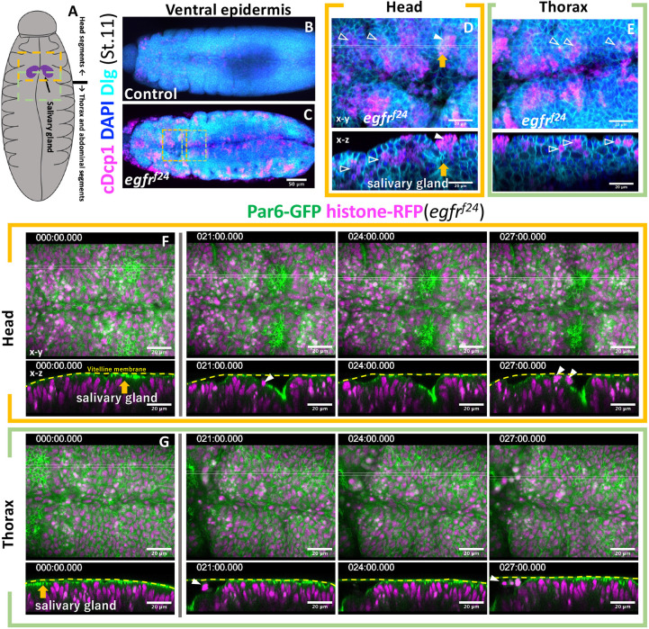
Fig. 4.
Apical cell extrusion at the site of vitelline membrane detachment. (A) Schematic of the ventral view of stage 11 embryos. (B-E) Active caspase staining (cDcp1) of control (B) and EGFR mutant (C) embryos. Parts of the head and thoracic regions of C (dotted box) are enlarged in D and E. Unfilled arrowheads, basally extruded cells; filled arrowheads, apically extruded cells; orange arrows, salivary gland invagination. (F,G) Time-lapse series of the ventral head and thorax imaged from two separate embryos, showing apical cell extrusion in the invaginating salivary gland primordia (orange arrows and white arrowheads). Other parts maintained tight vitelline membrane contact at this stage. Times are in minutes.

