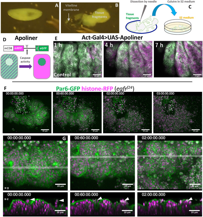
Fig. 5.
Rapid collapse of EGFR tissue fragments in explant cultures. (A-C) The procedure of embryo dissection. (D) Apoliner caspase activity detector. (E-G) Time-lapse imaging of cultured tissue fragments. (E) Apoliner activity in a control tissue fragment. (F) An EGFR mutant tissue fragment undergoing rapid disintegration. (G) The early phase of EGFR mutant tissue culture, showing frequent apical cell extrusion (arrowheads). Imaging started within 1 h of dissection (time is h:min:s).

