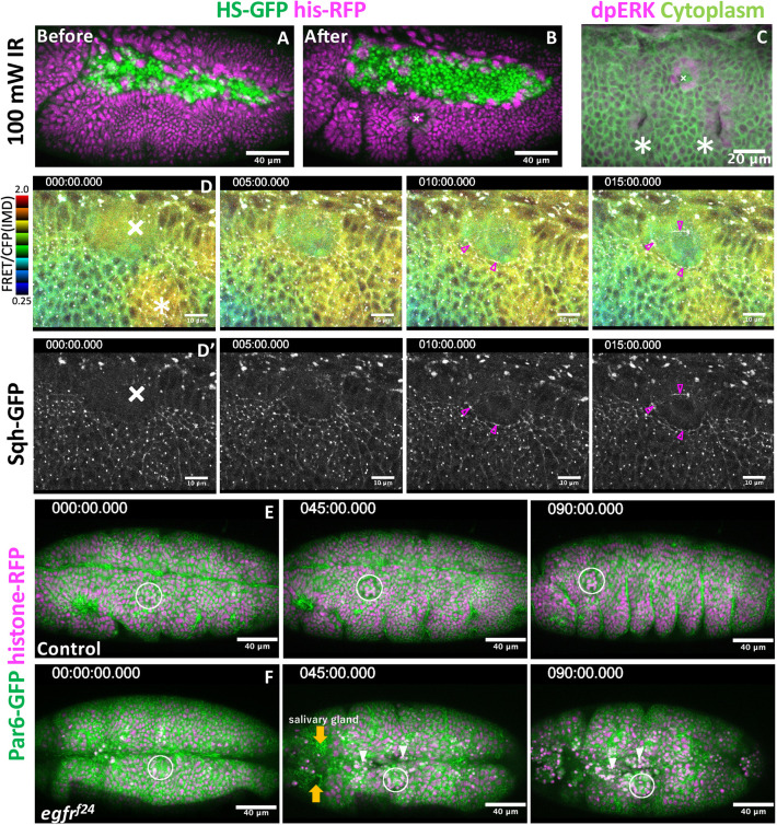
Fig. 6.
EGFR signaling is required for tissue repair. (A,B) Tissue wounds treated with infrared laser illumination. (C) ERK activation in the vicinity of the wound region. Crosses, wound site; asterisks, tracheal placode. (D,D′) Simultaneous recording of ERK activity (FRET probe, D) and myosin activity (D′). Magenta arrowheads indicate myosin accumulation around wound region. (E,F) Laser wound damage is limited locally in the control group (E). Tissue disintegration spreads from the wound site in EGFR mutants (F). White circles indicate the wound site. Orange arrows indicate invagination of the salivary gland. White arrowheads indicate apical cell extrusion around wound region. Times are in minutes.

