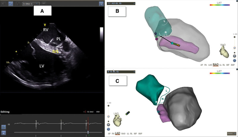
Figure 3.
Implantation of the left bundle branch lead under intracardiac echocardiography (ICE) guidance. A, Pacing lead (PL) implanted in the left ventricular (LV) endocardium through the interventricular septum (IVS) under ICE guidance. B and C, Three-dimensional model of LV and aorta (AO) reconstructed using ICE. The green part denotes the aorta, L denotes the left coronary aortic cusp (LCC), N denotes the noncoronary aortic cusp (NCC), and R denotes the right coronary aortic cusp (RCC). The pink part indicates the IVS, and the gray part indicates the LV. LAO indicates left anterior oblique view; LAT, local activation time; and RAO, right anterior oblique view.

