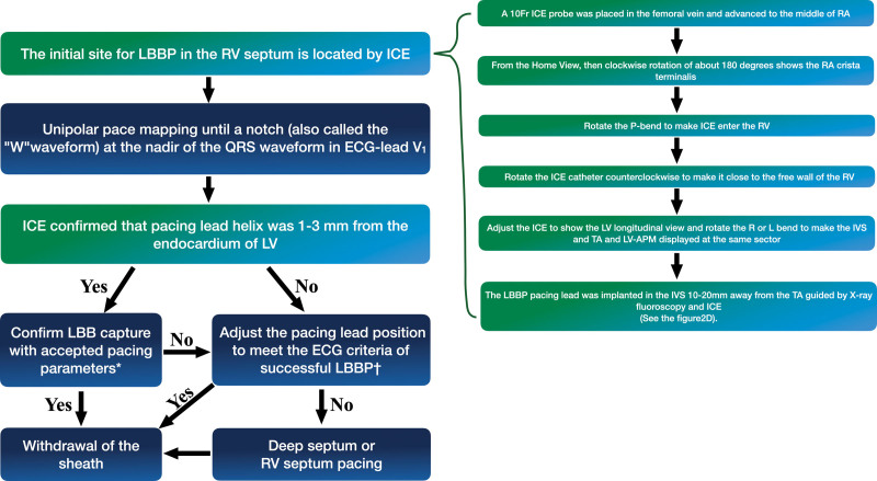
Figure 5.
Flowchart of the procedure for intracardiac echocardiography (ICE)–guided left bundle branch pacing (LBBP). *Confirm LBB capture with accepted pacing parameters: (1) pace the morphology of the right bundle branch block pattern; (2) record the LBB potential from the LBBP lead; (3) stimulus-peak left ventricular (LV) activation time (LVAT) shortens abruptly with increasing output and remains the shortest and constant at low and high outputs. †The method of adjustment of the pacing lead position involves the following aspects: (1) the pacing lead helix was closer to the endocardium of the LV monitored using ICE, (2) the pacing lead is positioned perpendicular to the IVS and enters the endocardium of the LV, as monitored using ICE, (3) under ICE guidance, the pacing lead was kept away from the scar area of the IVS. (4) The LBBP lead was implanted around the original site under the guidance of ICE. APM indicates anterior papillary muscle; RA, right atrium; RV, right ventricular; and TA, tricuspid annulus.

