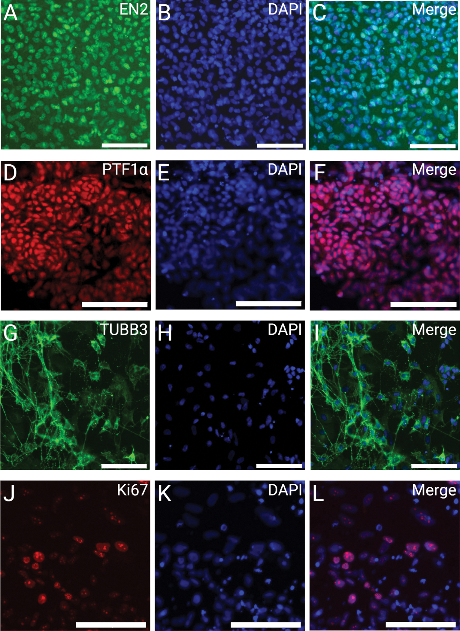
Figure 3: Immunofluorescent labeling of 2D cerebellar cells at day 35.

Cells are fixed with 4% PFA at day 35 and are nuclear stained with DAPI and immunolabeled for (A) EN2 (green), (B) DAPI (blue), (C) EN2-DAPI merged; (D) PTF1α (red), (E) DAPI (blue), (F) PTF1α-DAPI merged; (G) TUBB3 (green), (H) DAPI (blue), (I) TUBB3-DAPI merged; and (J) Ki67 (red), (K) DAPI (blue), (L) Ki67-DAPI merged. Scale bars: 100 μm.
