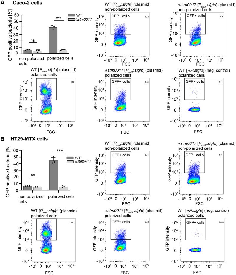
Fig 3. Quantification of chiA expressing S. Typhimurium subpopulations in contact with polarized intestinal epithelial cells.
The indicated S. Typhimurium fluorescence reporter strains were incubated for 3 h in contact with polarized and non-polarized Caco-2 (A) or HT-29 MTX cells (B). The bacteria were harvested and analyzed by flow cytometry to quantitate the subpopulations of GFP-expressing bacteria. The dot plots identify the gate that were set to determine GFP positive (GFP+) bacterial cells. The non-GFP expressing S. Typhimurium strain WT ΔP:sfgfp in contact with polarized IEC was used to evaluate background fluorescence. The bar graphs show the average percentages ± standard deviation of GFP positive cells derived from three independent experiments. Statistical analyses were implemented with ordinary one-way ANOVA and Bonferroni´s multiple comparisons test P>0.05: ns; P≤0.05: *; P≤0.01: **; P≤0.001: ***.

