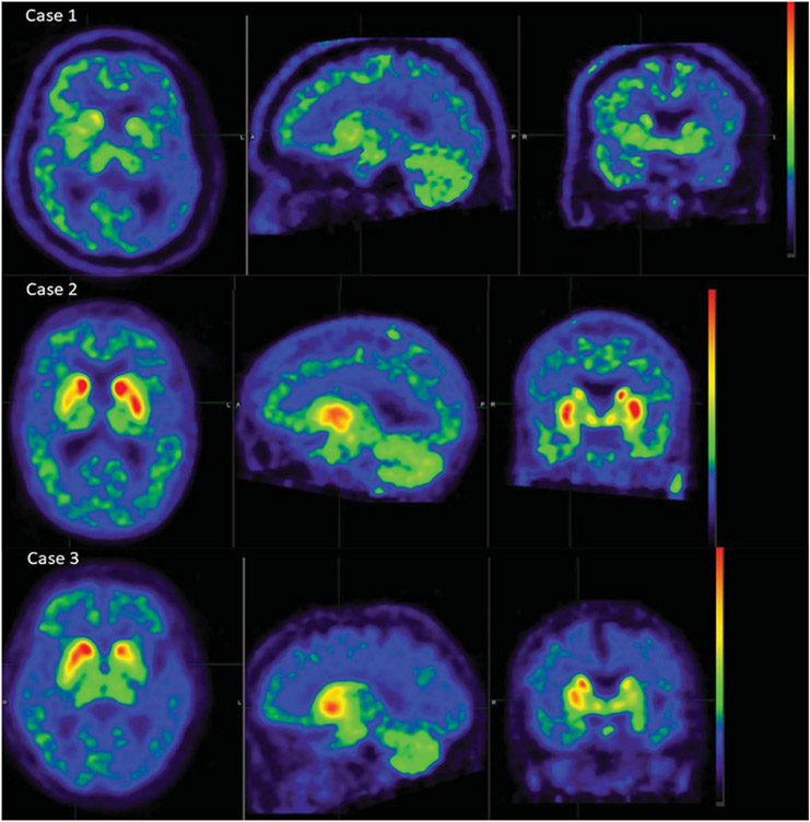
Fig. 1.
AV-133 PET images for the three studied cases. Case 1 (top) was read as having bilateral dopaminergic depletion in putamen and caudate by all 5 study readers. Case 2 (middle) was read as normal, without dopaminergic depletion in caudate or putamen, by all 5 readers. Case 3 (bottom) was read by all 5 readers as having a decreased signal in the left putamen, while 3/5 noted a decrease in right putamen and one indicated the decreases were asymmetrical, left > right; all 5 readers reported no decrease in left or right caudate nuclei.

