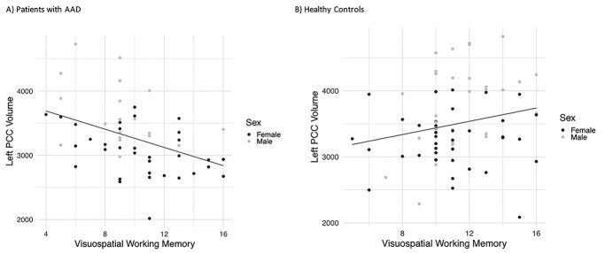
Fig. 2.
Associations between brain structure and visuospatial working memory. There was a significant interaction between group (patient or control) and volume of the left posterior cingulate cortex (PCC), and performance on a visuospatial working memory test (Span Board forward) (B = −0.003, 1 = 0.014). Post hoc tests revealed that in individuals with AAD, those with larger volume of the left PCC performed worse on the task (B = −0.003, P = 0.016), while in healthy controls, those with larger volumes performed better (B = 0.001, P = 0.048).

