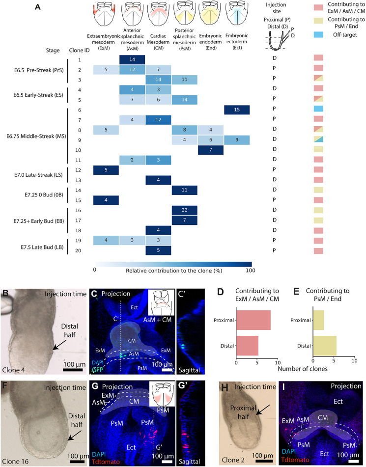
Fig. 3.
TAT-Cre microinjection allows prospective clonal analysis of nascent mesoderm. (A) Contribution of dose 1/2 TAT-Cre-induced clones. Squares contain cell counts (x-axis, clone; y-axis, location. n=19 embryos, eight litters). (B-C′) Injected embryo (B) and its resulting clone (C). C′ shows a sagittal plane of C. (D,E) Distal and proximal injections contributing to ExM, AsM and CM (n=14 embryos) (D) and to the PsM and End (n=8 embryos) (E). (F-G′) Injected embryo(F) and its resulting clone (G). G′ shows a sagittal plane of G. (H,I) Injected embryo (H) and its contribution (I) to embryonic and extra-embryonic compartments (n=2 embryos). Cardiac mesoderm is highlighted with white shading and embryonic compartments annotated following abbreviations in A.

