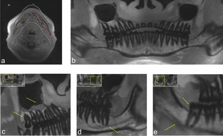
Figure 4.
OPG (orthopantomogram) - like MRI reconstructions (MR-OPGs) from a 3D ultrashort echo time (UTE) prototype sequence for bone and teeth imaging. (a) The planning of the orthopantomogram-like MR images. (b) Overview image of a UTE MR-OPG. (c) Visualization of the positional relationship of the maxillary third molar in the first quadrant and the maxillary sinus, with a retention cyst. (d) Visualization of the course of the hypointense inferior alveolar nerve (IAN) within the inferior alveolar canal through the mandible. (e) Shows the positional relationship between the mandibular third molar and the IAN in UTE MR-OPG.

