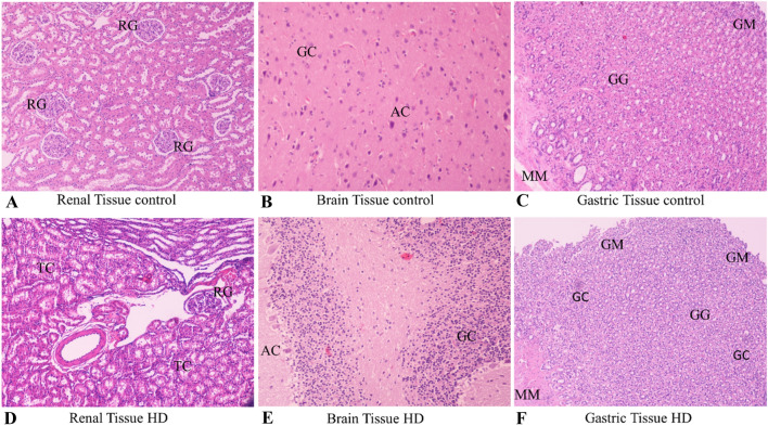
Fig. 2.
Slides of renal, brain and gastric tissue stained with eosin hematoxylin of control and high dose subjects. Magnification × 200. A Renal tissue control × 200. RG Renal glomerulus. Normal shape and number of renal glomeruli with normal cellularity level. B Brain tissue control × 200, AC astrocytes, GC glial cells. Normal number of glial cells and astrocytes. C Gastric tissue control × 200 GM Gastric mucosa, GG Gastric glands, MM Muscularis mucosae with normal architecture with no distortion of the mucosal integrity and no signs of inflammation. D Renal tissue high dose (HD) × 200. TC Tubular cells. Severe numerical reduction in cellularity with intense growth in tubular cell aggregates forming renal tubules molds. E Brain tissue high dose (HD) × 200, AC astrocytes, GC glial cells. Severe numerical reduction of glial and astrocyte cells. F Gastric tissue high dose (HD) × 200 GM Gastric mucosa, GG Gastric glands, GC granulocytes, MM Muscularis mucosae with severe distortion of mucosal architecture and necrotic lesions of mucosal foci along with mucosal degeneration

