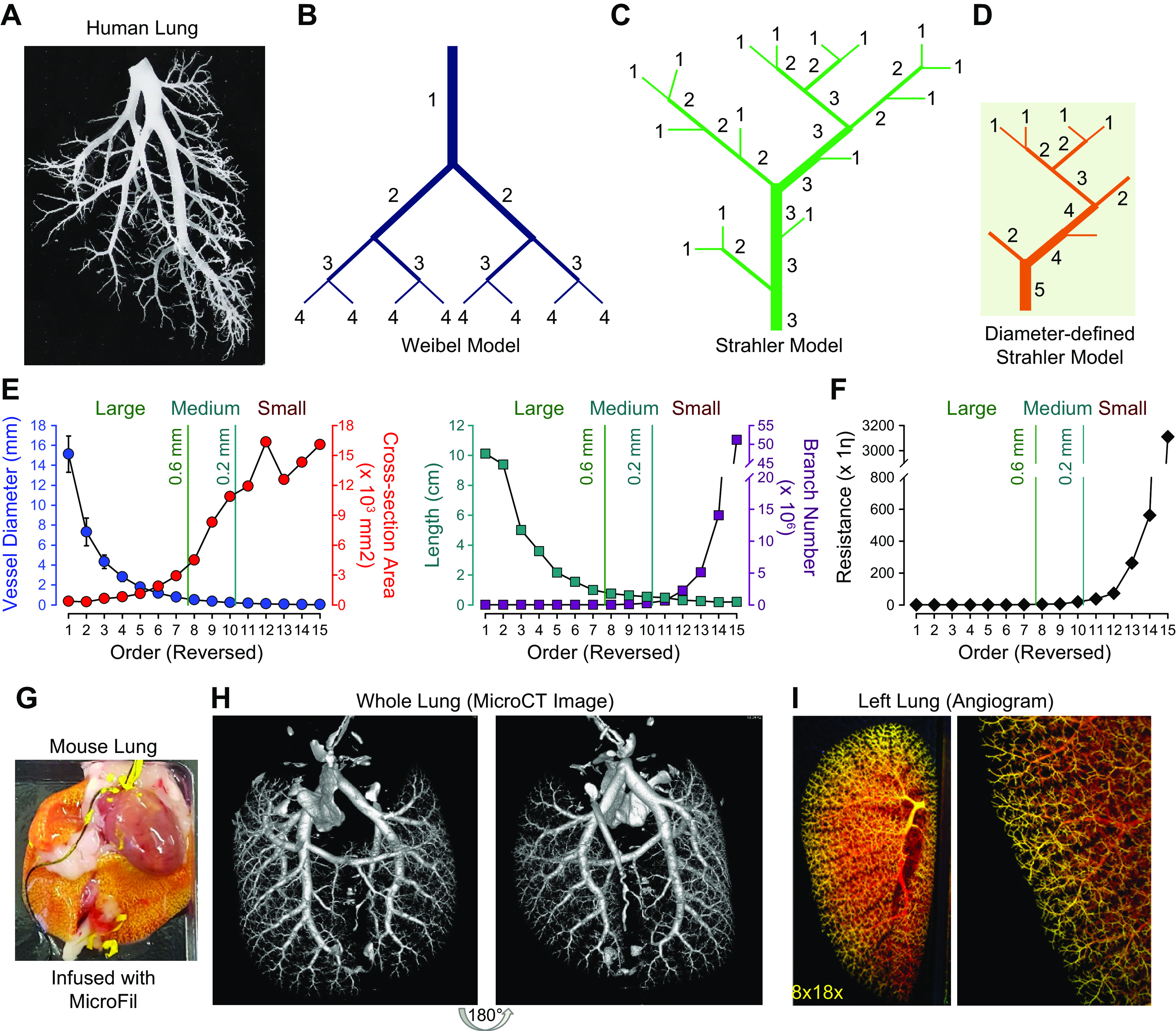
FIGURE 3.

A: typical cast of a small segment of the arterial tree in human lungs shows the complex structure of the vascular tree. B–D: the 3 proposed schemes describing this complex structure are illustrated by the Weibel model (B), the Strahler model (C), and the diameter-defined Strahler system (D). Generation numbers are indicated on each branch. Note that in the Weibel model the largest vessel is designated as a vessel of generation 1. After each bifurcation, the generation number of the offsprings is increased by 1. The exact opposite is true in the Strahler model and the diameter-defined Strahler system, in which the smallest noncapillary blood vessels are defined as order 1. In the Strahler model, when 2 vessels of the same order meet, the order number of the confluent vessel is increased. When 2 vessels of different orders meet, the order number of the confluent vessels remains the same as the larger of the 2. In the diameter-defined Strahler system, when 2 vessels of different order and diameter meet one another, the order number of the confluent vessel is increased only if its diameter is larger than either of the 2 segments by a certain amount. Otherwise, the order number of the confluent segment is not increased. E: lumen diameter (closed circles in blue; left) and length (closed squares in dark cyan; right) of each segment of pulmonary arteries and distribution of total cross-sectional area (closed circles in red; left) and number of branches (closed squares in dark red; right) of all segments of each order of pulmonary arteries in human lungs. Diameter and length of each of the individual pulmonary arterial branches decline exponentially from orders 1 to 15. The number of pulmonary arterial branches increases exponentially from orders 1 to 15. F: distribution of vascular resistance in different orders of pulmonary arteries showing that resistance increases exponentially from order 1 [the largest pulmonary artery (PA)] to order 15 (the smallest PA). Reproduced from Huang et al. (36). The vessels are subjectively classified into large (diameter > 0.6 mm), medium-sized (diameter = 0.6–0.2 mm), and small (diameter < 0.2 mm) based on their arterial diameter. G–I: MICROFIL-filled mouse lung via the right ventricle (RV) (G), high-resolution computerized tomography (CT) scan image of the mouse lung (H), and ex vivo angiogram of mouse lung (I) show peripheral pulmonary vascular branches.η, viscosity.
