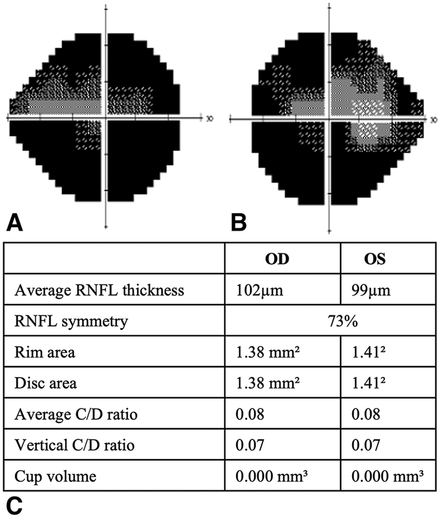
Figure 1.

Right(A) and left (A) HVF, corresponding RNFL thickness map(C) for a 20v/o male with severe visual field defects and papilledema
RNFL: Retinal Nerve Fiber Layer

Right(A) and left (A) HVF, corresponding RNFL thickness map(C) for a 20v/o male with severe visual field defects and papilledema
RNFL: Retinal Nerve Fiber Layer