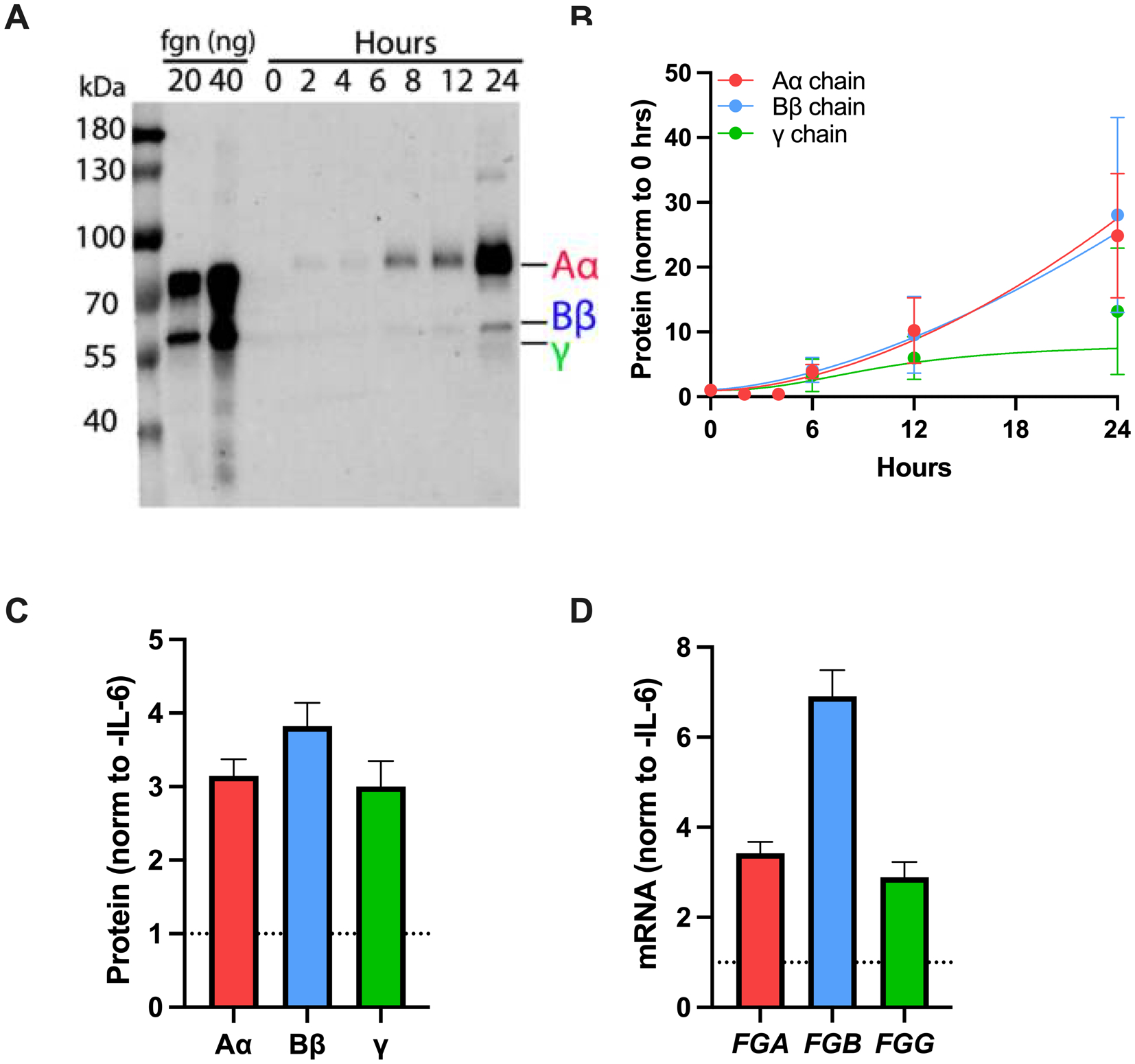
Figure 2. HepG2 cells are an experimental model of fibrinogen expression.

(A) Media was collected from HepG2 cells cultured in the absence or presence of IL-6 for 24 hours. Proteins were separated by reducing SDS-PAGE, transferred to polyvinylidene difluoride membrane, and probed with polyclonal rabbit anti-human fibrinogen and IR-DYE 800CW goat anti-rabbit antibodies. (B) Fibrinogen in the media was quantified by densitometry; symbols indicate mean ± standard error of the mean. (C-D) Effect of IL-6 (50 ng/mL) on fibrinogen (C) protein and (D) mRNA (N=4–6, bars indicate mean + standard error of the mean).
