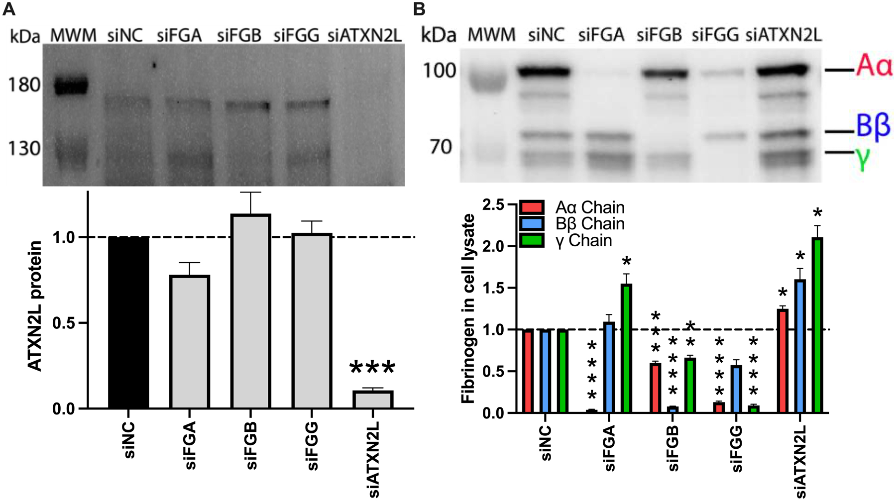
Figure 7. Loss of ATXN2L increases fibrinogen protein in HepG2 cell lysates.

HepG2 cells were transfected with siRNAs against FGA, FGB, FGG, or ATXN2L in the presence of IL-6. (A) ATXN2L protein was visualized by immunoblot and quantified by densitometry (upper band). (B) Fibrinogen was visualized in cell lysates by immunoblot and quantified by densitometry. All data were normalized to treatment with control siRNA (siNC), N=3–4 for each condition, bars indicate mean+standard error of the mean; *P < 0.05, **P < 0.01, ***P < 0.001, ****P < 0.0001
