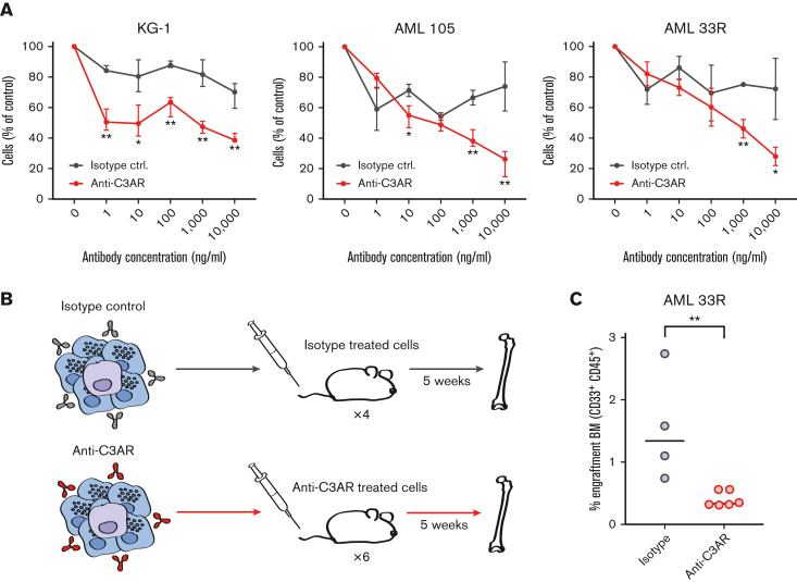
Figure 7.
C3AR can be specifically targeted with anti-C3AR antibodies. (A) ADCC assay on AML cell line KG-1 and 2 primary NPM1-mutated AML samples. The red line shows the percentage of AML cells after overnight incubation with a polyclonal anti-C3AR antibody and NK cells, compared with control without antibody. The black line shows the isotype-treated cells under the same experimental conditions. Error bars show mean with standard deviation. (B) Schematic illustration of the ADCC in vivo repopulation assay; residual cells from an ex vivo ADCC experiment on a primary AML sample were transplanted into immunodeficient NSG mice, which were sacrificed after 5 weeks. AML cells are depicted in purple and NK cells in blue. (C) Leukemic engraftment after ADCC, as the percentage of CD45+CD33+ human cells in the BM of mice that received transplantation with isotype-treated or anti-C3AR–treated AML cells, as in panel B. Antibody (10 μg/mL) was used. Line at median. ∗P ≤ .05 and ∗∗P ≤ .01 using a Student t test in panel A and a nonparametric Mann-Whitney U test in panel C.

