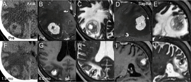
Figure 4. Computed tomography and magnetic resonance images before and after stereotactic radiosurgery.
The images show axial images (A-C, F-H); sagittal images (D, E, I, J); computed tomography images (A, F); CE-T1-WI (B, D, G, I); T2-WI (C, E, H, J); before SRS (A-E); at 1.9 months after SRS (F); and at 6.0 months (G-J).
(A-J) All images were co-registered and are shown in the same magnification (A-J) and coordinates (A-F). (F) At 1.9 months after SRS, considerable shrinkage of the BM was observed along with modest alleviation of the perilesional edema and mass effect. (G-J) At 6.0 months, marked shrinkage of the BM and mitigation of the peritumoral edema were observed along with extrication from the mass effect.
CE: contrast-enhanced; WI: weighted image; SRS: stereotactic radiosurgery; BM: brain metastasis

