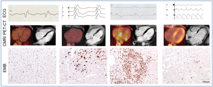
Figure 2. Examples of diagnostic findings in patients with CS.

Fulfillment of current diagnostic criteria may vary because of different points in time of presentation (subclinical, active, quiescent, or burned‐out disease). All of the following findings are associated with patients with cardiac sarcoidosis and positive endomyocardial biopsy. Upper row (left to right): incomplete right bundle branch block, right bundle branch block, atrioventricular block III, and ventricular tachycardia. Middle row (left to right): no FDG uptake in PET‐CT and no late gadolinium enhancement in CMR, no FDG uptake in PET‐CT and late gadolinium enhancement in CMR, FDG uptake in PET‐CT and no late gadolinium enhancement in CMR, and FDG uptake in PET‐CT and late gadolinium enhancement in CMR. Bottom row (left to right): normal finding (sampling error), CD3+ T lymphocytes in lymphocytic myocarditis, CD3+ lymphocytes in noncaseating granuloma, and isolated CD3+ T lymphocytes and fibrosis; all images ×200. CMR indicates cardiovascular magnetic resonance; CS, cardiac sarcoidosis; EMB, endomyocardial biopsy; FDG, F18‐fluorodeoxyglucose; and PET‐CT, positron‐emission tomography‐computed tomography.
