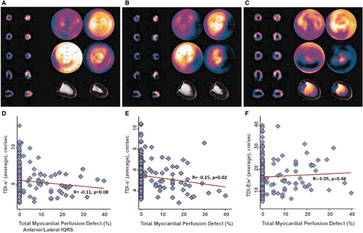
Figure 2. Myocardial perfusion defects in (A) non‐fQRS, (B) inferior fQRS, and (C) anterior/lateral fQRS with SPECT imaging.

D through F, Correlation between total myocardial perfusion defect and TDI‐derived myocardial early relaxation (TDI‐e′) velocity, systolic (TDI‐s′) velocities, and LV filling pressure E/e′. fQRS indicates fragmented QRS; LV, left ventricular; SPECT, myocardial perfusion single‐photon emission computed tomography; and TDI, tissue Doppler imaging.
