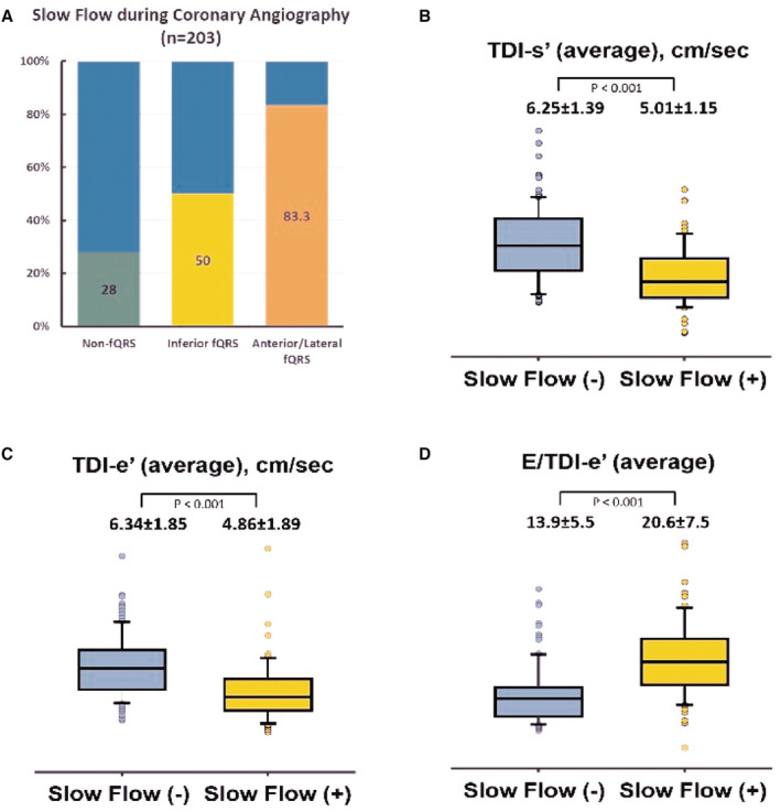
Figure 3. Association between the presence of coronary slow flow and cardiac function.

A, The percentage of coronary slow flow during coronary angiography among 203 out of 634 (32.0%) non‐CAD study participants. B through D, Comparisons of the TDI‐s′, TDI‐e′, and average E/e′ in normal versus slow coronary flow non‐CAD study participants. CAD indicates coronary artery disease; fQRS, fragmented QRS; and TDI, tissue Doppler imaging.
