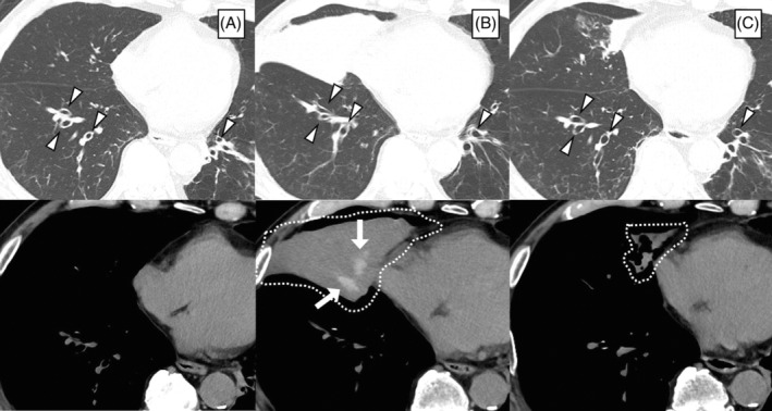
FIGURE 1.

Computed tomography images of the chest (A) before the administration of mepolizumab, (B) when developing allergic bronchopulmonary aspergillosis during mepolizumab treatment, and (C) 3 months after initiating treatment with tezepelumab. Arrowheads indicate central bronchiectasis. High‐attenuation mucus plugs in the right middle lobe of the lung (white arrows), accompanied by atelectasis (dotted lines), were attenuated after the initiation of tezepelumab therapy
