Fig. 2.
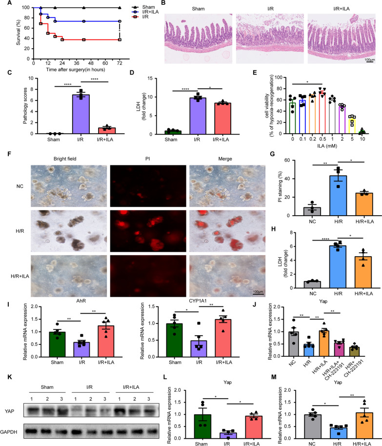
ILA improved the survival rate and inhibited apoptosis of enterocytes under I/R and H/R injury. A Survival rate between sham, I/R and I/R + ILA groups (n = 16/group). B-C H&E staining (B) and quantification (C) of the histopathology changes of intestinal tissue sections (n = 3/group). D Relative serum LDH levels in mice under intestinal I/R injury (n = 4/group). E The effect of different ILA concentration on organoids’ cell viability under H/R injury (n = 5/group). F Representative images of morphological changes and PI staining in organoids. G Quantification of PI staining (n = 3/group). H Relative LDH levels in the organoids culture supernatant (n = 3–4/group). I AhR and CYP1A1 mRNA levels in intestinal tissues (n = 5/group). J YAP mRNA levels in organoids (n = 5/group). K Western blot for the protein expression of YAP in intestinal tissues (3 representative cases in each group). L YAP mRNA levels in intestinal tissues (n = 4/group). M YAP mRNA levels in organoids (n = 5/group). Results are presented as mean ± SEM. The statistical tests employed included: two-tailed log-rank test in (A), two-tailed student’s t-test in C–E, G–I, L and M and one-way ANOVA followed by the Tukey test for multiple comparisons in J. * p < 0.05, ** p < 0.01, **** p < 0 .0001

