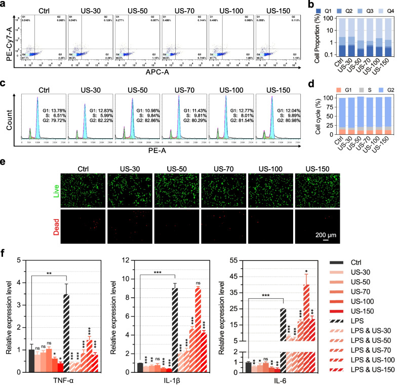
Fig. 3.
Effects of LIPUS stimulation on the proliferation and inflammation of C28/I2 cells. a Flow cytometry analysis for cell apoptosis of C28/I2 cells stimulated with LIPUS. b Quantification data for apoptosis rate of C28/I2 cells. c Flow cytometry analysis for cell cycle distribution of C28/I2 cells. d Quantification data for the cell cycle of C28/I2 cells. e Representative live-dead staining images of C28/I2 stimulated with LIPUS; scale bar: 200 μm. f qPCR analysis of the expression of TNF-α, IL-1β, and IL-6 in C28/I2 cells, after LPS treatment (to produce inflammation) and LIPUS stimulation. *p < 0.05, **p < 0.01, ***p < 0.001 (n = 3)

