Figure 2.

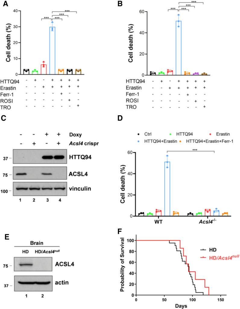
The role of ACSL4 in mHTT-induced ferroptosis and the life span of the HD mice. (A) Cell death assay. HTTQ94 tet-on HT-22 cells preincubated with 0.5 μg/mL doxycycline for 4 h were treated with 1 μM erastin for 12 h in the presence or absence of 2 μM ferrostatin-1 (Ferr-1) or ACSL4 inhibitors (10 μM rosiglitazone [ROSI] and 10 μM troglitazone [TRO]). (B) Cell death assay. HTTQ94 tet-on SK-N-BE(2)C cells preincubated with 0.5 μg/mL doxycycline for 16 h were treated with 40 μM erastin for 32 h in the presence or absence of 2 μM ferrostatin-1 (Ferr-1), 10 μM rosiglitazone (ROSI), and 10 μM troglitazone(TRO). (C) Western blot analysis for ACSL4 and HTTQ94 in HTTQ94 tet-on SK-N-BE(2)C control and Acsl4-Crispr cells treated with 0.5 μg/mL doxycycline for 16 h. (D) Cell death assay. HTTQ94 tet-on SK-N-BE(2)C control Crispr and Acsl4-Crispr cells were preincubated with 0.5 μg/mL doxycycline for 16 h and then treated with 40 μM erastin for 32 h with/without 2 μM Ferr-1. (E) Western blot analysis for ACSL4 from the cerebral cortex tissues of HD and HD/Acsl4-null mice. (F) Kaplan–Meier survival curves of HD (n = 21 independent mice) and HD/Acsl4-null (n = 7 independent mice) mice. Cell deaths were calculated from three replicates. Data shown in A, B, and D are the means ± SD. P-values were derived from two-tailed unpaired t-test. (***) P ≤ 0.001. In F, P-value (HD vs. HD/Acsl4-null) was calculated using log-rank Mantel–Cox test. P = 0.1356.
