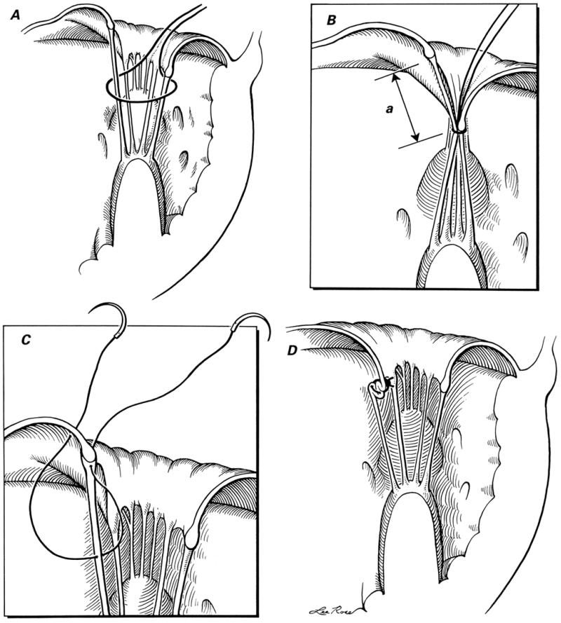
Fig. 2 Technique of chordal shortening: A) a silk suture is passed around both the chorda to be shortened and the opposite (posterior cusp) chorda; B) after the left ventricle has been filled with saline solution, the silk suture is tightened so that the anterior mitral leaflet's degree of prolapse can be assessed and the length of chorda to be shortened (a) can be determined; C) after exposure of the chorda with a traction suture (not shown), a 5-0 polypropylene double-armed suture is taken, and the needle is passed through the chorda and then through the edge of the anterior mitral leaflet close to its point of attachment; and D) the polypropylene suture is tied, shortening the chorda at cusp level.
(Illustrations by Lee Rose. Reproduced with permission. From Texas Heart Institute Journal 1992;19:47–50.)
