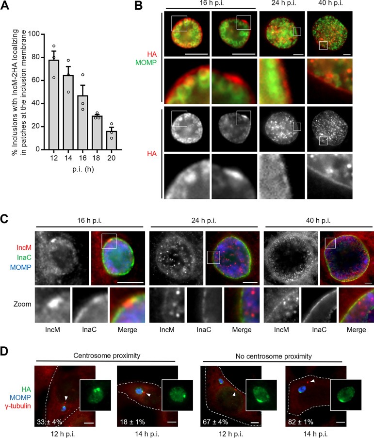
FIG 1.
IncM concentrates transiently at patches in the inclusion membrane. HeLa cells were infected at a multiplicity of infection of 0.3 for the indicated times with C. trachomatis strain incM::aadA harboring a plasmid encoding IncM with a C-terminal 2HA epitope tag (pIncM-2HA) (A, B, and D) or with the L2/434 strain (C). The infected cells were fixed with methanol, immunolabeled with anti-C. trachomatis MOMP (green) and anti-HA (red) antibodies (A and B), anti-MOMP (blue), anti-IncM (red), and anti-InaC (green) antibodies (C), or anti-MOMP (blue), anti-HA (green), and γ-tubulin (centrosomes; red) (D) antibodies and analyzed by fluorescence microcopy. (A) Enumeration of the number of inclusions showing IncM-2HA localizing in patches at the inclusion membrane at different times postinfection (p.i.). (B) Representative images of the localization of IncM-2HA during C. trachomatis infection of HeLa cells at different times p.i. (C) Images of the localization of endogenous IncM during C. trachomatis infection of HeLa cells at different times p.i. (D) Illustrative images and enumeration of the number of IncM-2HA patches at the inclusion membrane at 12 and 14 h p.i. that localized within ~2 μm from the centrosome (centrosome proximity) or not (no centrosome proximity); arrowheads point to the centrosomes. For panels A and D, values represent means ± standard errors of the mean, n = 3, ≥40 cells (A) or ≥20 cells (D) per experiment. Scale bars, 5 μm.

