FIG 3.
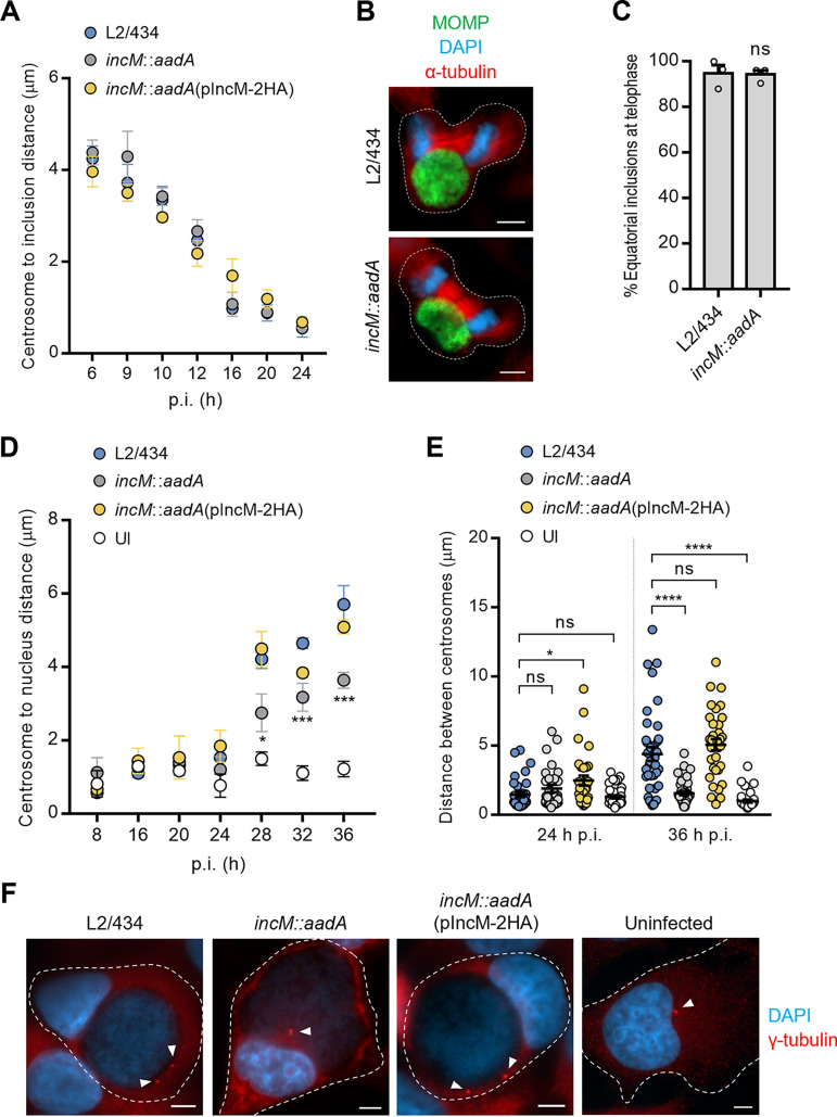
IncM contributes to host cell centrosome positioning in infected cells. HeLa cells were infected at a multiplicity of infection of 0.1 with C. trachomatis strains L2/434, incM::aadA, or incM::aadA harboring a plasmid encoding IncM with a C-terminal 2HA epitope tag (pIncM-2HA), as indicated. Cells were fixed with methanol at the indicated times postinfection (p.i.) (A, D, and E) or only at 24 h p.i. (B and C), immunolabeled with anti-C. trachomatis MOMP and γ-tubulin (centrosomes) antibodies, and stained for nuclei with DAPI (blue). (A) The distance of the centrosomes to the closest point of the inclusion was measured by fluorescence microscopy using Fiji (75). (B) Representative infected cells show the localization of the inclusion during telophase. Scale bars, 5 μm. (C) Quantification of the position of the inclusion in telophase cells relative to the division plane (equatorial versus polar). (D) The distance of the centrosomes to the closest point in the nucleus was measured using Fiji (75). (E) The distance between centrosomes was measured using Fiji (75). (F) Representative uninfected (UI) and infected cells at 36 h p.i showing centrosome positioning; arrowheads point to the centrosomes. Scale bars, 5 μm. Values represent means ± standard errors of the means, n = 3, ≥30 cells per experiment. P values were obtained by a two-tailed t test (C) or by one-way ANOVA and Dunnett’s post hoc test analysis relative to cells infected by the L2/434 strain (D and E). ns, not significant; *, P < 0.05; ***, P < 0.001; ****, P < 0.0001.

