Fig. 5.
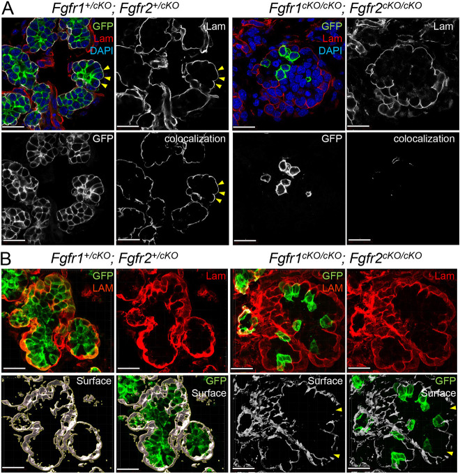
Cell-basement membrane interactions in Fgfr1/2 compound mutants. (A) Cell-basement membrane (BM) interactions in the submandibular gland (SMG) were analyzed for compound Fgfr1/2 compound mutants (n=24) at E15.5 by generating 3D colocalization maps. The BM was marked by laminin (Lam). K14 lineage epithelial cells expressed GFP. High-resolution confocal imaging followed by 3D rendering was used to detect colocalization between GFP of laminin and colocalization maps were generated. Control cells formed robust cell-BM contacts (yellow arrowheads). Several GFP+, Fgfr1cKO/cKO; Fgfr2cKO/cKO cells did not interact with the BM. Scale bars: 20 µm. (B) The integrity of the BM in compound Fgfr1/2 mutants was analyzed. 3D surfaces around laminin were generated from high-resolution 3D rendered confocal images. Compound Fgfr1cKO/cKO; Fgfr2cKO/cKO mutants showed discontinuity along the laminin surfaces (yellow arrowheads). Scale bars: 20 µm.

