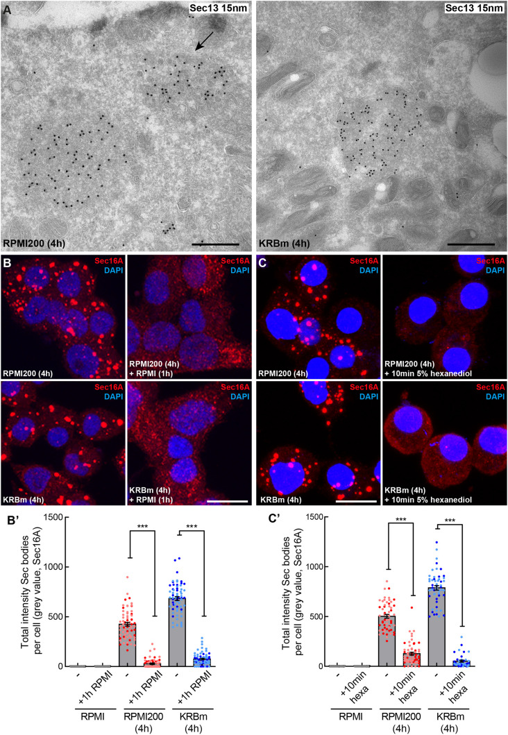
Fig. 3.
Sec16A-positive structures in stressed INS-1 cells are Sec bodies. (A,A′) Visualization of endogenous Sec13 (15 nm PAG) by immunoelectron microscopy in ultrathin frozen sections of INS-1 cells incubated in RPMI200 and KRBm for 4 h. Sec13 is concentrated in structures (Sec bodies) that are slightly denser than the surrounding cytoplasm, are round, are not sealed in a lipid bilayer and are in close proximity to the ER. Arrow points to a remaining ERES that have not remodeled upon stress. (B,B′) IF visualization of Sec16A in INS-1 cells after 4 h in RPMI200 and KRBm followed by 1 h in RPMI (B). Note that Sec body dissolution is complete within 1 h of stress removal and that Sec16A is localized again at ERES. Quantification in B′; N=2 experiments, n=64–69 cells. (C,C′) IF visualization of Sec16A in INS-1 cells treated with 5% hexanediol (hexa) for 10 min after incubation in RPMI200 or KRBm for 4 h (C). Quantification in C′; N=2 experiments, n=40–60 cells. Scale bars: 500 nm (A), 10 μm (B) and (C). Error bars are s.e.m. (B′,C′). ***P<0.001 (Mann–Whitney test).

