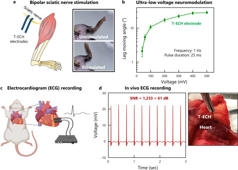
Fig. 5. In vivo bioelectronic device functionalities of T-ECH.
a Schematic illustration and image of bipolar sciatic nerve stimulation with T-ECH electrodes on the sciatic nerve. b Leg moving angle was measured for various stimulation voltages. Values represent the mean and the standard deviation (n = 9 devices examined with each mouse). The frequency was set to 1 Hz and the pulse duration was 25 ms. c Schematic illustration of in vivo ECG monitoring using T-ECH electrodes. Created with BioRender.com. d High-quality recording of ECG signals. SNR = 1122 (61 dB).

