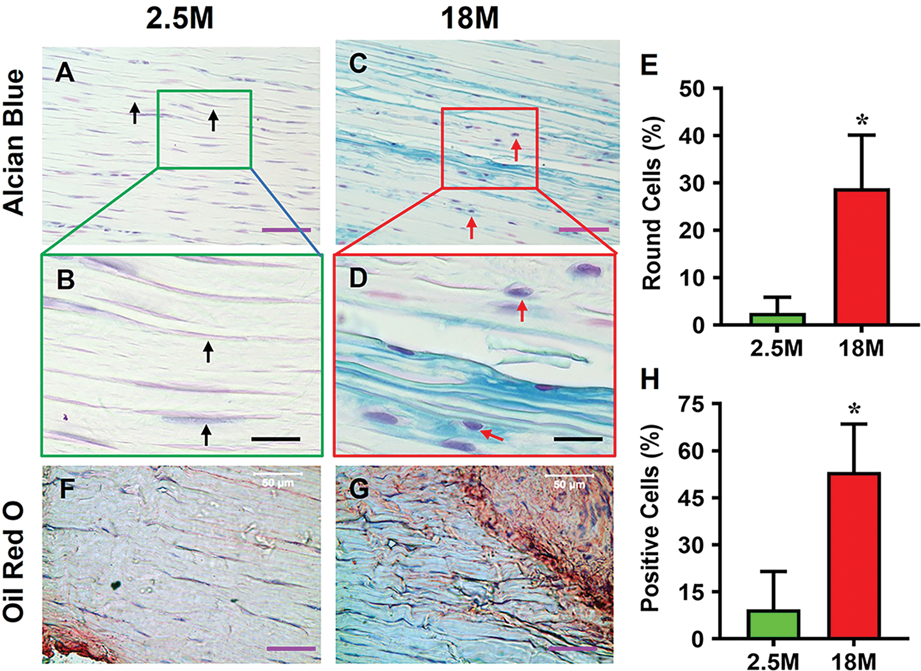
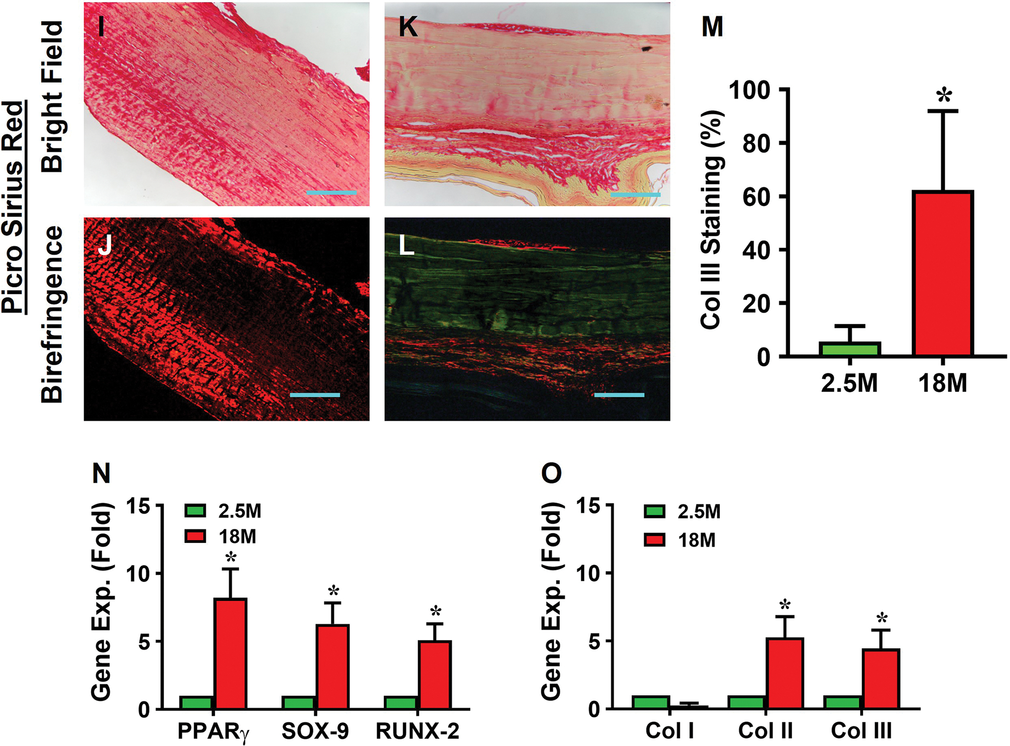
Fig. 1. Aging tendon exhibits degenerative changes.


Histochemical staining for proteoglycans (PG) by Alcian blue shows minimal staining for PG in young tendon, and the cells exhibit an elongated morphology (A, B, black arrows). In contrast, aging tendon shows robust presence of PG along with round shaped cells (C, D, red arrows). Semi-quantification shows a significantly higher number of round cells in aging tendon compared to young tendon, with more than 28% round cells in aging tendon vs 2% cells in young tendon I. Similarly, young tendon does not show the presence of lipids by Oil Red O staining (F), while aging tendon has extensive lipid staining (G, red area). Aging tendon has significantly greater lipid staining compared to young tendon, with 53% staining in aging vs 9% in young tendon as shown by semi-quantification (H). Picro Sirius Red staining shows that young tendon under light microscope is formed by strong collagen fibers (red in I), while aging tendon under light microscope is formed by loose collagen fibers (yellow in K). Polarized light microscopy results indicate that the thick collagen fibers in young tendon are formed by collagen type I (red/yellow in J), while the loose collagen fibers in aging tendon are formed by collagen type III (green in L). Semi-quantification shows 62% of collagen fibers in aging tendon are collagen III, but 5.7% of collagen fibers in young tendon are collagen III (M). Gene analysis by qRT-PCR shows significantly decreased expression of collagen I (Col I) for tendon-related gene marker, and increased expression of non-tenocyte-related genes, PPARγ for adipocytes, SOX-9 and collagen II (Col II) for chondrocytes, Runx-2 for osteocytes, and collagen III (Col III) for scar tissue in aging tendon compared to those in young tendon (N, O). Purple bars: 50 μm; Black bars: 12.5 μm; Blue bars: 100 μm. *p < 0.05 (aging compared to young).
