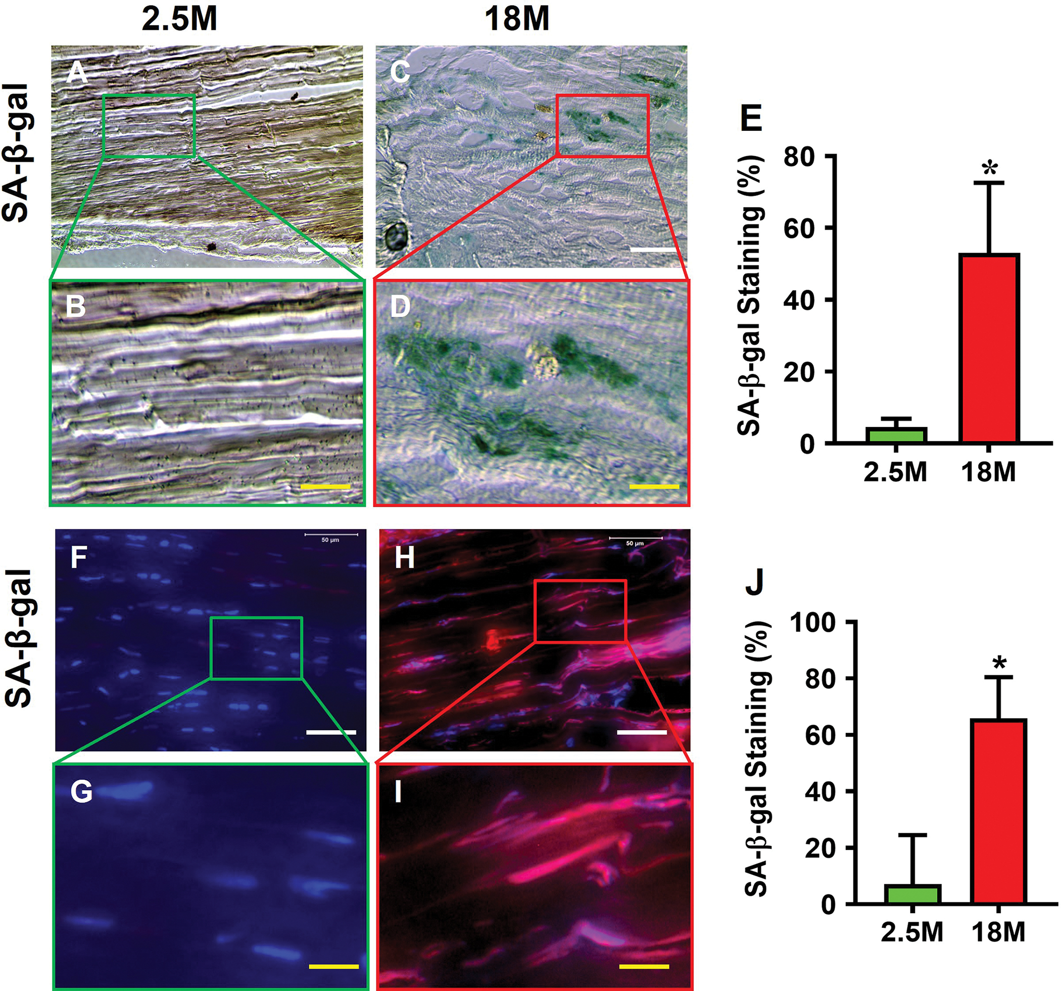
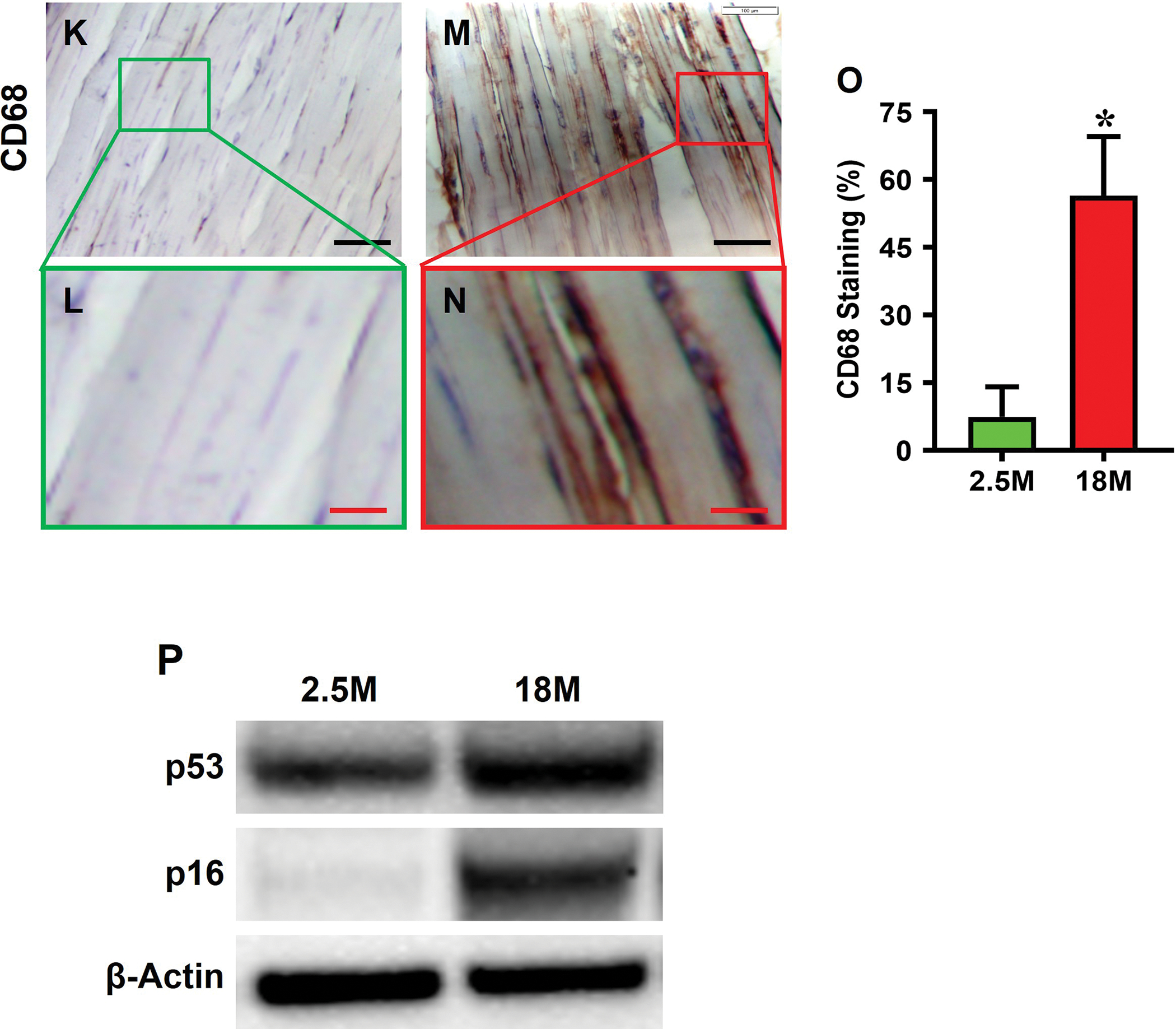
Fig. 2. Senescent cells are present in aging tendon.


Histochemical (top panel) and immunostaining (bottom panel) show that few cells are positively stained with SA-β-gal in young tendon (A, B, F, G), but abundant staining is evident in aging tendon (green in C, D and red in H, I), which are confirmed by semi-quantification (E, J). Immunostaining for CD68 on young tendon tissue section shows minimal staining (K, L), but aging tendon tissue section exhibits abundant positive staining (M, N, brown), which are confirmed by semi-quantification showing 56.5% positive staining in aging vs 7.5% in young tendon (O). Western blot results show that the expression of senescent cell markers p53 and p16 are greatly increased in aging tendon compared to young tendon, which shows nearly no expression of p16 (P). Black bars: 100 μm; White bars: 50 μm; Red bars: 25 μm; Yellow bars: 12.5 μm; *p < 0.01 (aging compared to young).
