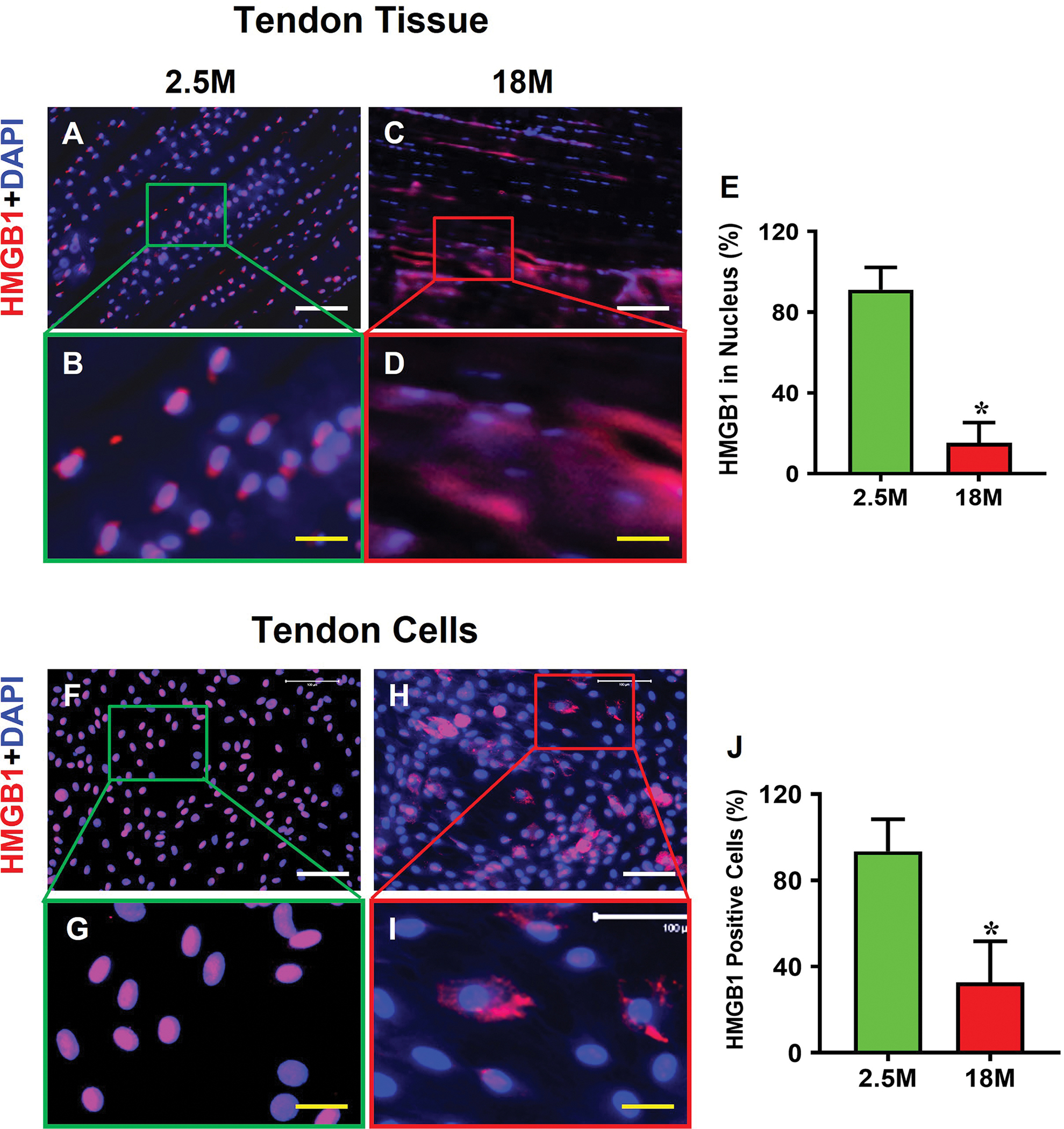
Fig. 3. HMGB1 is translocated from the nucleus to cytoplasm and extracellular matrix in aging tendon.

Immunofluorescence analysis on mouse tendon tissue sections shows that HMGB1 is present within the cell nuclei in young tendon (A, B). In contrast, HMGB1 in aging tendon is translocated to cytoplasm (C, D). Semi-quantification results of tendon tissue sections indicate that 91% of the cells in young tendon have HMGB1 within the nuclei, but only 15% of the cells in aging tendon have HMGB1 in the cell nuclei (E). Similarly, isolated cells cultured from young tendon harbor HMGB1 within the cell nuclei (F, G), whereas the aging cells show HMGB1 staining in the cytoplasm (H, I). Semi-quantification confirms the results showing that 93% of the cells in young tendon cells have HMGB1 in the nuclei with 33% in aging tendon cell nuclei (J). White bars: 100 μm; Yellow bars: 25 μm, *p < 0.01 (aging compared to young).
