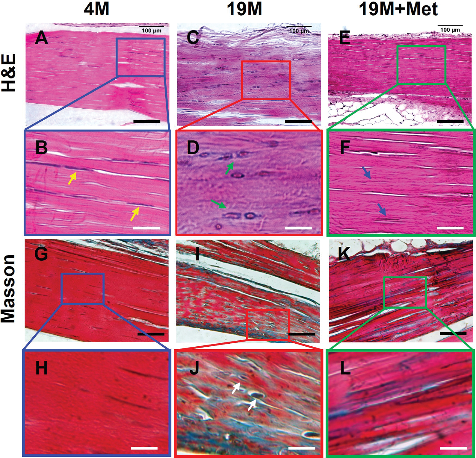
Fig. 9. IP injection of Met decreases degenerative changes in aging tendon.

H&E staining on young mouse (4M) tendon tissue section shows that the cells are elongated in shape (B, yellow arrows), whereas cells in aging tendon (19M), are round shaped (D, green arrows). However, Met injection decreases the number of round shape cells in aging tendons (F, blue arrows). Masson trichrome staining on young tendon tissue sections shows that it is formed by dense collagen fibers all stained red (G, H), but the aging tendon has some blue staining interspersed across the tendon section, indicating loose collagen fibers with tendon cells (J, white arrows). However, IP injection of Met for 8 weeks decreases the presence of loose collagen fibers in aging tendon (K, L). Black bars: 100 μm; White bars: 25 μm.
