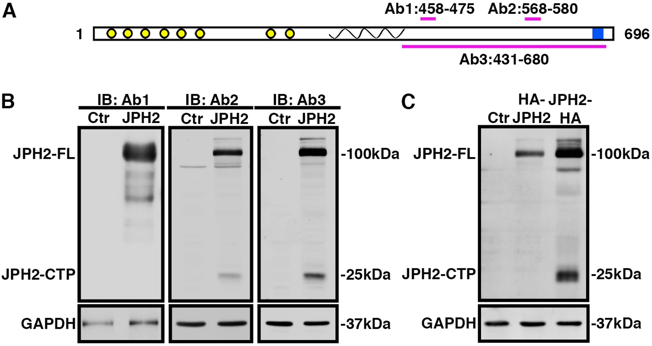
Fig. 1. JPH2 is cleaved at its C-terminus.

A) Cartoon of JPH2 protein with N-terminal MORN domains (yellow), α-helix (spiral), and C-terminal transmembrane domain (blue). Antibody epitopes are labeled Ab1, Ab2 and Ab3. B) Western blots using three different JPH2 antibodies revealing expression of full-length (FL) JPH2 and a 25-kDa C-terminal peptide in lysates of HEK293 cells expressing either JPH2 or pcDNA3.1 vector control (Ctr). C) Western blots using HA antibody revealing expression of JPH2-FL and/or JPH2-CTP in lysates of HEK293 cells that were not transfected (Ctr; lane 1), expressing HA-JPH2 (lane 2), or JPH2-HA (lane 3), respectively. All the experiments were performed at least 3 times to validate reproducibility.
