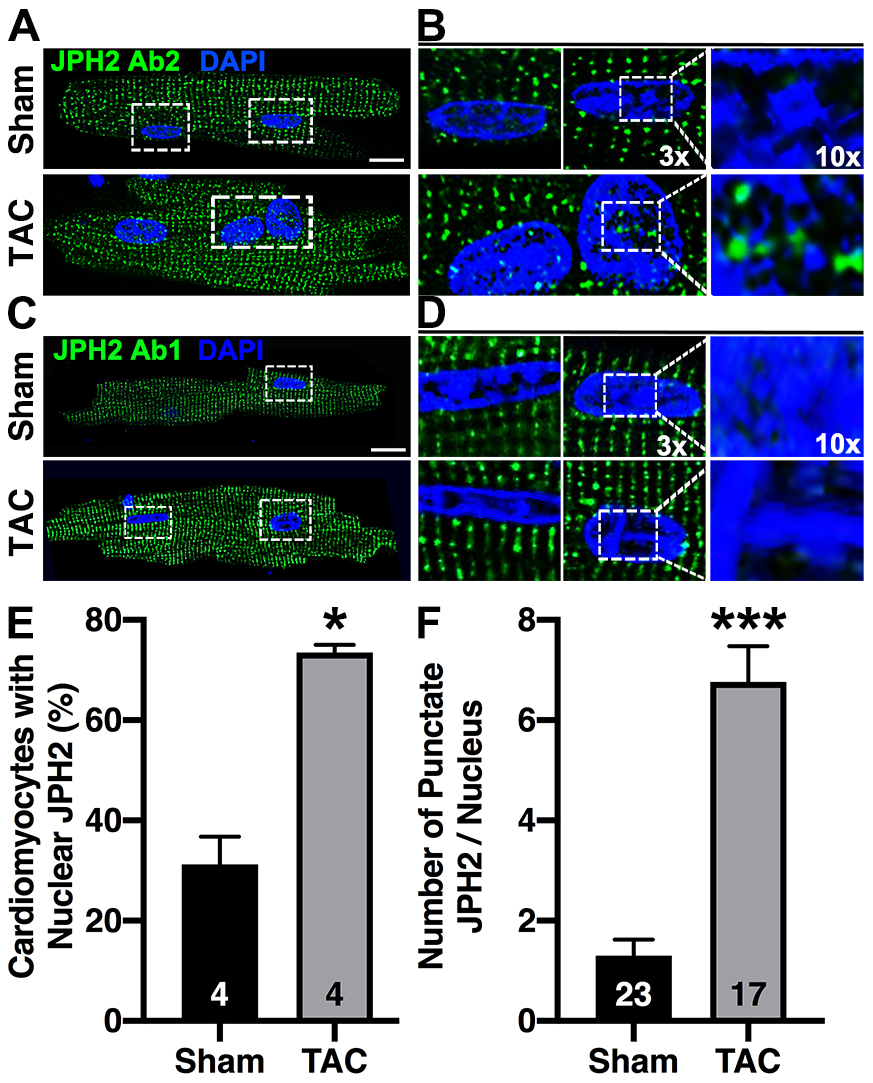
Fig. 6. JPH2-CTP translocates into the nucleus in myocytes from mice subjected to TAC.

Immunofluorescence of fixed ventricular cardiomyocytes isolated from mice subjected to TAC or a sham procedure following staining with A) JPH2 C-terminal antibody (Ab2); 3x and 10x zoom images are shown on the right (B). C) Staining with JPH2 Ab1 which is N-terminal to the CTP; 3x and 10x zoom images on the right (D). White dotted box indicates zoom region. Scale bar = 10μm. E) Quantification of the percent of cardiomyocytes with positive nuclear staining after TAC, N = number of mice. F) Quantification of average number of JPH2 puncta per nuclei after TAC-induced HF, N = number of nuclei. Data were analyzed using Mann-Whitney test (*P<0.05; **P<0.01; ***P<0.001). All the experiments were performed at least 3 times to validate reproducibility.
