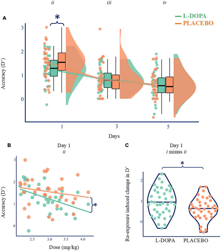
FIGURE 2.
Nocturnal dopamine dose-dependently modulates memory. (A) Paired comparisons for d’ across time. D’ was higher on Day 1 on placebo (orange; d’ mean ± standard deviation = 1.54, ±0.65) compared with L-DOPA (green; mean = 1.25 ± 0.59; Wilcoxon’ = 121, p = 0.004, BF10 = 16.6, Cohen’s δ = 0.46, n = 35), but there was no difference on Days 3 or 5 (p > 0.05). Boxplots show quartiles with kernel densities plotted to the right. *p < 0.05. (B) Higher L-DOPA dose during consolidation correlated with poorer Day 1 recall of single exposure items (List ii d’; Spearman’s ρ = –0.560, p < 0.001, n = 35) but no such relationship was found on the placebo night (ρ = –0.231, p = 0.180, orange). These two relationships were also different [r-to-z transform z = –2.554, p = 0.011, (Lee and Preacher, 2013)]. Lines of best fit presented for illustration purposes. *p < 0.05. (C) L-DOPA increased the relative benefit of re-exposed compared to other items (List i d’ minus List ii d’; Wilcoxon = 444, p = 0.034, BFio = 2.6, Cohen’s δ = 0.42, n = 35, SM9). Lines show maximum, median, and minimum values (horizontally) and kernel densities (vertically). *p < 0.05.

