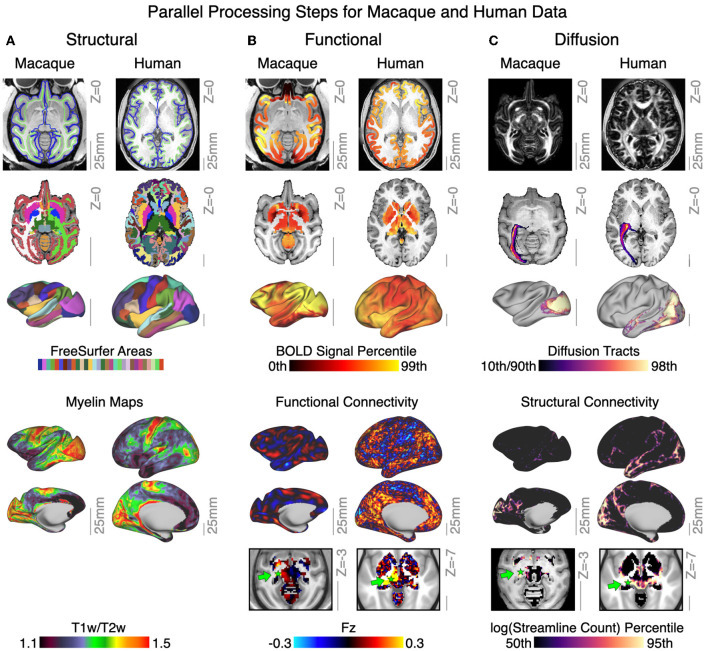
Figure 6.
QuNex enables neuroimaging workflows across different species. (A) Structural features for exemplar macaque and human data, including surface reconstructions and segmentation from FreeSurfer. Lower panel shows output myelin (T1w/T2w) maps. (B) Functional features for exemplar macaque and human showing BOLD signal mapped to both volume and surface. Lower panels show and resting-state functional connectivity seeded from the lateral geniculate nucleus of the thalamus (green arrow). (C) Diffusion features for exemplar macaque and human data, showing whole-brain fractional anistropy, and volume and surface terminations of the left optic radiation tract. Lower panels show the structural connectivity maps seeded from the lateral geniculate nucleus of the thalamus (green arrow). Gray scale reference bars in each panel are scaled to 25 mm.

