
Fig 4. Disruption of Sonic Hedgehog signaling impedes post-stroke neurogenesis.
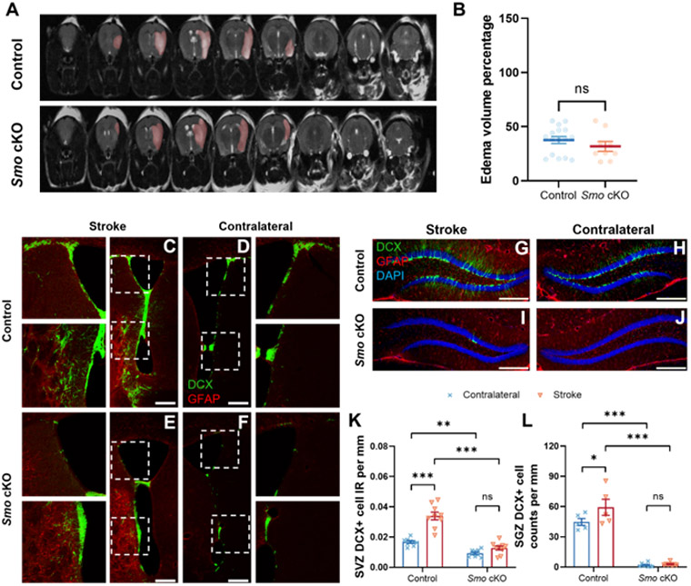
A T2-weighted MRI scan visualized infarction area following tMCAo stroke (highlighted by pink color). B Calculated infarction index showed statistical comparable infarct volume between Smo cKO and control mice (Control, n=16. Smo cKO, n=9, Students’ t-test). At one month after stroke, Immunostaining of DCX-labeled immature neurons (Green) and GFAP-labeled reactive astrocytes (Red) at SVZ region from control (C, D) or Smo cKO mice (E, F) at ipsilateral lesion side (C, E) and contralateral non-lesion side (D, F). SGZ region DCX-labeled immature neurons (Green) and GFAP-labeled reactive astrocytes (Red) from control (G, H) or Smo cKO mice (I, J) at ipsilateral lesion side (G, I) and contralateral non-lesion side (H, J). Quantification results showed significant increase of DCX+ cell IR at lesion side SVZ (K, Control: n=9. Smo cKO: n=9, 2way ANOVA with Tukey’s post-hoc test) or cell counts at lesion side SGZ (L, Control: n=5. Smo cKO: n=8, 2way ANOVA with Tukey’s post-hoc test) were observed in control stroke mice but not in Smo cKO mice. Scale bar= 250 μm. All data shown are Mean±SEM. * p<0.05, ** p<0.01, *** p<0.001. For statistical detail, see Table S1.