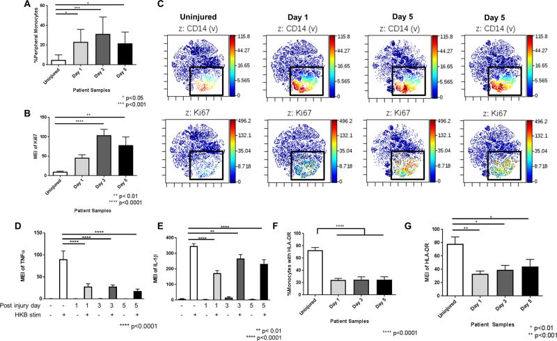
Figure 3. Circulating monocytes increase in percentage with increasing Ki-67 expression in trauma patients, but have reduced pro-inflammatory cytokine and HLA-DR expression.
A. Percentages of circulating monocytes in peripheral blood demonstrating that the percentage of circulating monocytes increases significantly at all time points after injury. B. Mean expression intensity (MEI) of Ki-67 expression in monocytes demonstrating a significant increase in Ki-67 expression at all time points after injury. C. viSNE plots illustrating the colocalization of CD14 and Ki67 staining of proliferating monocytes. Top panels show CD14 staining and bottom panels show Ki67 staining of same cell population. D. MEI of TNFα expression in monocytes in peripheral blood after stimulation with heat killed bacteria (HKB). Stimulated TNFα expression in monocytes decreased at all time points after injury. E. MEI of IL-1β in HKB-stimulated monocytes, showing a significant decrease in IL-1β expression at all time points after injury. F. Percentages of circulating HLA-DR+ monocytes from uninjured controls and trauma patients. The percentage of circulating monocytes that were HLA-DR+ was significantly reduced at all time points after injury. G. MEI of HLA-DR on monocytes is significantly decreased at all time points after injury. Statistics were performed using one-way ANOVA; results were considered significant if p < 0.05.

