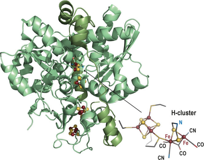
Figure 4.

Ribbon representation (light green) of the [FeFe]-hydrogenase crystal structure (PDB 1HFE(11)). [Fe4S4] clusters are represented as balls and sticks with the Fe and the S atoms colored in dark red and yellow, respectively. A zoom-in of the H-cluster is represented as balls and sticks for the iron and sulfide ions; the ligands are represented as sticks with N, C, O and S atoms colored in blue, gray, red and yellow, respectively.
