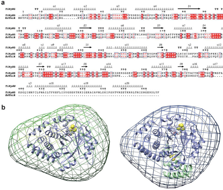
Figure 6.
(a) Sequence alignment of TiHydG and EcThiH primary structures. Red and white boxes indicate conserved and similar amino acids in the two proteins. TiHydG secondary structure elements are depicted on top. (b) SAXS-generated envelope for (left) a HydG mutant lacking the C-terminal domain, HydGΔ91 from Thermoanaerobacter tengcongensis (Tte), and (right) wild-type TteHydG.51 The TiHydG crystal structure27 without (left, blue ribbon representation) and with (right, blue and green ribbon representation) the C-terminal domain and auxiliary cluster (see main text) were placed in the envelope. [FeS] clusters are represented as van der Waals spheres.

