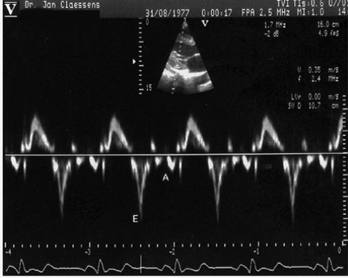
Fig. 4 Pulsed Doppler tissue imaging showing the peak early diastolic velocity (E) and peak atrial systolic velocity (A) of the LV posterior wall, with sample volume at the endocardial portion of the basal site of the LV posterior wall in the parasternal long-axis echocardiogram.
