Figure 1.
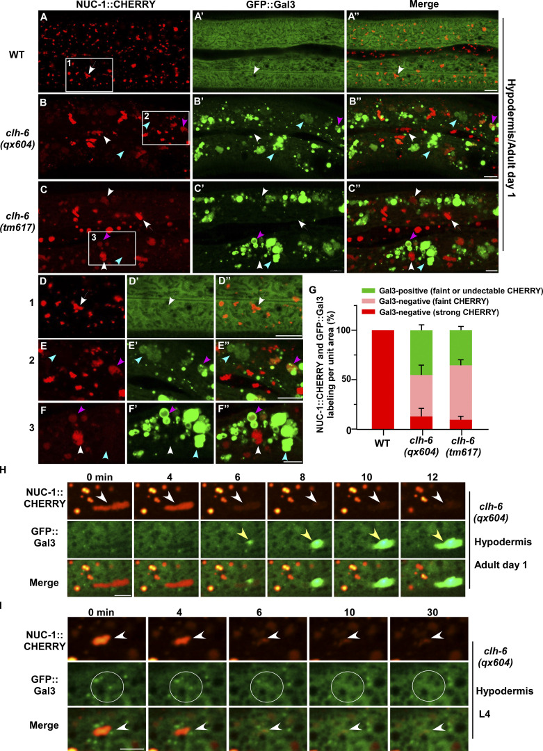
clh-6 mutants accumulate damaged lysosomes. (A–G) Confocal fluorescence images of the hypodermis in wild-type (A–A′′), clh-6(qx604) (B–B′′), and clh-6(tm617) (C–C′′) adults co-expressing NUC-1::CHERRY and GFP::Gal3. The boxed region in A–C is magnified in D–F. Intact lysosomes contain strong NUC-1::CHERRY fluorescence and are not labeled by GFP::Gal3 (white arrows). Damaged lysosomes have either faint NUC-1::CHERRY fluorescence with GFP::Gal3 labeling (purple arrowheads) or without GFP::Gal3 labeling (white arrowheads), or strong GFP::Gal3 fluorescence with undetectable NUC-1::CHERRY (blue arrowheads). Quantification is shown in G. At least 10 animals were scored in each strain and data are shown as mean ± SD. (H and I) Time-lapse images of lysosomes in clh-6(qx604) expressing GFP::Gal3 and NUC-1::CHERRY. “0” min represents the time point before NUC-1::CHERRY release (white arrowheads) and initial appearance of GFP::Gal3 (yellow arrowheads). Circles indicate the region where NUC-1::CHERRY leakage but not GFP::Gal3 enrichment was observed. Scale bars: 5 µm.

