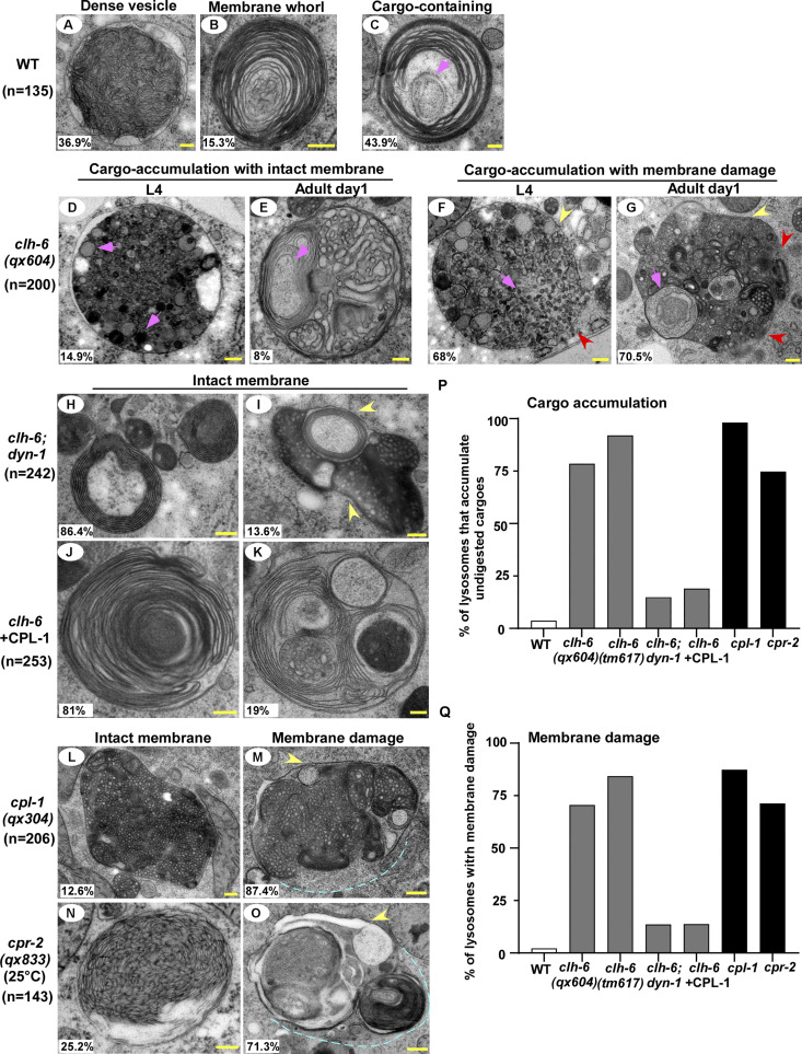
Figure 2.
Loss of clh-6 causes rupture of lysosome membranes. (A–O) Transmission electron micrographs of lysosomes in the hypodermis of wild type (A–C), clh-6(qx604) (D–G), clh-6;dyn-1 (H and I), clh-6 with CPL-1 overexpression (J and K), cpl-1(qx304) (L and M), and cpr-2(qx833) (N and O). Yellow arrowheads indicate lysosomal membranes, while red arrowheads and blue dashed lines designate areas that have no detectable membranes. Cargoes and granules are indicated by pink arrowheads. The percentage of lysosomes with the representative pattern is quantified and shown at the lower left corner in each panel. (P and Q) The percentage of lysosomes that accumulate undigested cargoes (P) or with membrane damage (Q) is quantified in the indicated stains. Scale bars: 200 nm.

