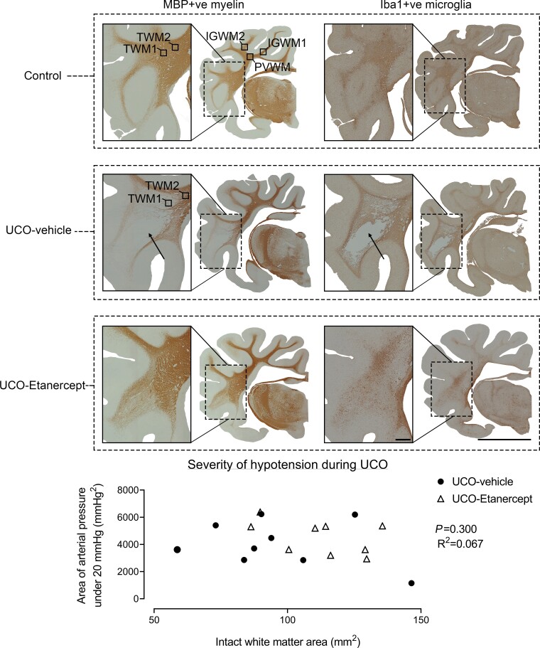
Figure 1.
Macroscopic WMI. Examples of coronal sections of the right hemisphere and enlargement of the temporal lobe after 21 days recovery from UCO labelled for MBP-positive myelin proteins (left column) and Iba-1-positive microglia (right column). Note the cystic white matter lesions observed in the temporal lobe in the UCO-vehicle group (arrows), and absence of cystic lesions in the control and UCO-Etanercept groups. Images were taken at ×2.5 magnification. The top left coronal section shows the five white matter regions assessed for microscopic analysis. Scale bar = 10 mm for the right hemisphere and 1 mm for enlarged sections. Bottom graph shows that no relationship was found between the severity of hypotension during UCO (assessed as the area of arterial pressure under 20 mmHg) and the intact white matter area.

