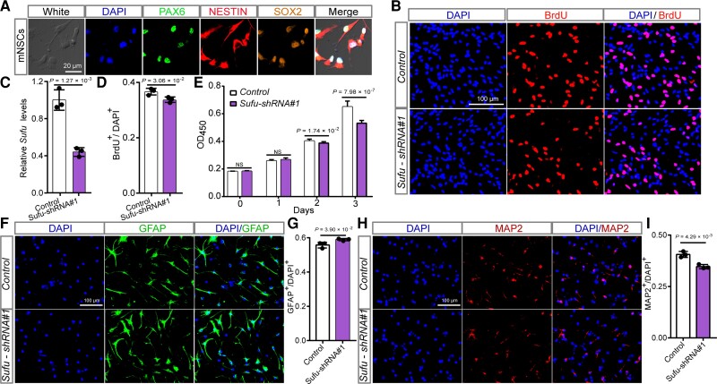
Figure 4.
Sufu knockdown inhibits proliferation and neurogenesis of mNSCs. (A) Immunofluorescence staining results of three well-characterized markers (SOX2, PAX6 and NESTIN) for neural stem cells confirmed the identity of the isolated mNSCs. (B) Immunofluorescence staining for BrdU incorporation assay. Red indicates BrdU-positive cells undergoing DNA amplification and DAPI was used to stain the nucleus (blue). (C) Sufu expression was efficiently knocked-down by the shRNA in mNSCs. (D) The quantification results of the BrdU incorporation assay. (E) The results of CCK-8 assay. Data were collected at 0, 1, 2 and 3 days after plating. (F) Immunofluorescence staining images for astrocyte cells (GFAP-positive cells) differentiated from mNSCs. (G) Quantification data for the ratio of GFAP-positive cells. Sufu knockdown led to a significant increase of GFAP-positive cells. (H) Immunofluorescence staining images for mature neurons (MAP2-positive cells) differentiated from mNSCs. (I) Quantification data for the percentage of MAP2-positive cells in total cells. Sufu knockdown resulted in a significant decrease of MAP-positive cells. n = 3 for B–D, F–I; n = 9 for E. mNSCs, mouse neural stem cells. Unpaired two-tailed Student’s t-test; data are presented as mean ± SD. NS, not significant.

