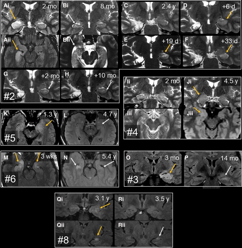
Figure 2.
Serial T2 or FLAIR MRI with time since disease manifestation. The images show the mediotemporal lobes with swelling and signal increase (orange arrows), HS (grey arrows) or normal findings. Each box contains images from one patient. Each letter marks one study time point; subscript numbers discriminate different orientations or planes. (A–H) Patient 2. [A(i and ii)] Two months, right-sided hippocampal volume and signal increase, amygdala unaffected. [B(i and ii)] Six months later, there is right-sided HS and still no amygdalar lesion. (C) Almost 2 years after right-sided selective amygdalohippocampectomy and start of prednisolone therapy, the patient is seizure-free and still has intact left mediotemporal structures. Prednisolone was discontinued. (D) A few days later, the patient developed left temporal status epilepticus, and the left hippocampus showed increased volume and T2 signal. (F and G) Close-meshed follow-up MRIs over the following 2 months revealed how this resulted in left HS. (H) Final stage. (I and J) Patient 4. [I(i and ii)] Two months after epilepsy onset, mediotemporal structures were normal; 3.8 years after onset, there was swelling and signal increase of the right amygdala (not shown). [J(i and ii)] These changes were still present 9 months later, that is, 4.5 years after onset. In this case, the amygdala but not the hippocampus was affected; immediately after J, the right amygdala was resected because a glioma was suspected. (K and L) Patient 5. (K) The first available MRI at 1.3 years after onset displays the left amygdalar and hippocampal volume and signal increase. (L) On the next available MRI study, there is already hippocampal atrophy (better visible on the not shown coronal sections). (M and N) Patient 6. (M) Three weeks after onset, there is bilateral swelling and signal increase of both hippocampal heads and amygdalae. (N) Five years later, the MRI reveals bilateral HS. (O and P) Patient 3. (O) At 3 months, the left hippocampus (and amygdala, not shown) are swollen and hyperintense. (P) Fourteen months after onset, left-sided HS has evolved. (Q and R) Patient 8. [Q(i and ii)] On the earliest available images taken 3.1 years after onset, the left amygdala and hippocampus are swollen and hyperintense. [R(i and ii)] Four months later, the left amygdala has returned to normal [R(i)], while the hippocampus has become atrophic with still increased signal, that is, HS [R(ii)]. d = days; mo = months; wks = weeks; y = years.

