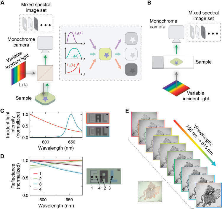
Fig. 4. Spectral imaging with highly multicolored arrays.
Schematic depicting the concept behind microscale (A) reflected-light and (B) transmitted-light spectral imaging using variable incident illumination and a monochrome camera. (C) Example of different incident light spectra and the corresponding reflected-light microscope images. Scale bar, 40 μm. (D) Reconstructed reflectance spectra at different spots on the sample shown in the optical micrograph, using the emitters in table S4. Scale bar, 40 μm. (E) Spectral data cube for a human tissue sample imaged using a transmitted-light imaging setup. Scale bars, 100 μm.

